Read Trigger Point Therapy for Myofascial Pain Online
Authors: L.M.T. L.Ac. Donna Finando
Trigger Point Therapy for Myofascial Pain (34 page)

S
OLEUS
Proximal attachment:
Posterior aspect of the head of the fibula and the proximal one-third of the posterior fibula and middle one-third of the tibia.
Distal attachment:
Via the tendocalcaneus (Achilles tendon) to the posterior surface of the calcaneus, with gastrocnemius.
Action:
Plantar flexion of the foot; assists inversion; contributes to knee stability; provides ankle stability.
Palpation:
To identify soleus, locate the following structures:
- Tendocalcaneus (Achilles tendon)âThe thickest and strongest tendon in the body, the tendocalcaneus is the common tendon of insertion for the gastrocnemius and soleus muscles. This tendon is palpable from the lower one-third of the calf to the calcaneus.
Soleus lies deep to gastrocnemius. Palpate soleus on the distal one-half of the lower leg, lateral and distal to the lateral head of gastrocnemius and medial and distal to the medial head of gastrocnemius.
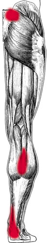
Soleus pain pattern
Pain pattern:
Heel pain, tenderness, restricted dorsiflexion; walking uphill or up stairs may be difficult. Trigger points in the proximal aspect of the muscle radiate to the posterior calf; distal trigger points radiate to the posterior aspect of the heel, possibly including its plantar surface and the distal aspect of the Achilles tendon; pain may be experienced at the sacroiliac joint on the same side.
Causative or perpetuating factors:
Chronic overload due to excessive plantar flexion; sudden overload due to misstep; poor circulation in the legs.
Satellite trigger points:
Gastrocnemius, homolateral quadriceps.
Affected organ system:
Cardiovascular system.
Associated zones, meridians, and points:
Dorsal zone; Foot Tai Yang Bladder meridian, Foot Shao Yin Kidney meridian; BL 59, KI 7, KI 9.
Stretch exercises:
- Place the ball of the foot on a step or curb and allow the heel of the foot to drop below the level of the step. Keep the knee bent as you stretch the calf. Hold this position for a count of twenty-five to thirty.
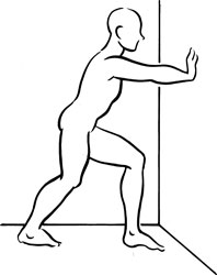
- Stand approximately 12 inches away from a wall, the hands placed on the wall at chest level. Place the leg to be stretched approximately 18 inches behind the other, keeping the toes of both feet facing the wall and the feet hip-width apart. Bend both knees to stretch soleus. Hold this position for a count of twenty-five to thirty.
Strengthening exercise:
Standing and holding on to a wall or chair for balance, raise up onto the ball of the foot, bringing the heel well off the floor. Hold this position for a count of five. Slowly return the heel to the floor. Repeat ten to twelve times.

Stretch exercise 1: Soleus
Stretch exercise 2: Soleus

Tibialis posterior and trigger point

Tibialis posterior pain pattern
T
IBIALIS
P
OSTERIOR
Proximal attachment:
Proximal two-thirds of the posterior surfaces of the tibia and the fibula and the interosseus membrane.
Distal attachment:
Passing behind the medial malleolus to attach to most of the bones that form the arch of the foot: the navicular, each cuneiform, and the cuboid; the calcaneus; and metatarsals 2, 3, and 4.
Action:
Inverts and adducts (supinates) the free foot; assists in plantar flexion. Prevents excessive pronation of the foot during walking; prevents excessive weight bearing on the medial foot; distributes weight evenly along the heads of the metatarsals, helping to shift the weight laterally.
Palpation:
Tibialis posterior is the deepest muscle of the calf, lying between the interosseus membrane anteriorally and the soleus posteriorally. Along with flexor digitorum longus and flexor hallucis longus it comprises the deep posterior compartment of the leg. Due to the depth of its placement on the leg this muscle cannot be palpated directly; however, tenderness can be elicited through deep palpation. Identify the posterior border of the tibia on the posteromedial surface of the leg. Palpate the proximal half of the lower leg, partially displacing the soleus posteriorly, to identify the posterior surface of the tibia through the overlying soleus.
Pain pattern:
Tibialis posterior is rarely involved in isolation from other calf muscles. Pain radiates over the Achilles tendon above the heel; additional pain may cover the midcalf, the heel, and the plantar surface of the foot and toes. Pain is experienced in the sole of the foot when walking or running, particularly on uneven surfaces.
Causative or perpetuating factors:
Jogging on uneven surfaces such as crowned roads; hypermobility of the midfoot; badly worn footwear that does not protect against eversion and rocking of the foot.
Satellite trigger points:
Flexor digitorum longus, flexor hallucis longus, and the peroneal muscles.
Affected organ systems:
Genitourinary system.
Associated zones, meridians, and points:
Dorsal zone; Foot Tai Yang Bladder meridian; BL 55, 56, and 57.
Stretch exercise:
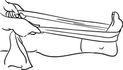
Stretch this muscle by dorsiflexing and everting the foot. Sitting with the legs extended in front of you, place a belt or towel around the midfoot of the leg to be stretched. Pull the band toward you, stretching the posterior calf. Pull with slightly greater force on the outside of the foot, which will allow the lateral side of the foot to be everted. Hold this position for a count of fifteen to twenty. Repeat three to five times.
Strengthening exercise:
Due to the nature of this muscle, strengthening exercises are generally not necessary.
Stretch exercise:Tibialis posterior

Tibialis anterior and trigger point
T
IBIALIS
A
NTERIOR
Proximal attachment:
Lateral condyle of the tibia and the upper one-half to two-thirds of the lateral surface of the body of the tibia, interosseus membrane, and surrounding fascia.
Distal attachment:
Medial and plantar surfaces of the medial cuneiform bone and the base of the first metatarsal.
Action:
Dorsiflexion and supination (inversion) of the foot. Helps to maintain standing balance, prevents foot slap at heel strike, and helps foot clear floor at swing phase. Vigorously active during most sports activities, including jogging, running, sprinting, and two-legged jumps. Helps maintain standing balance.
Palpation:
Tibialis anterior, peroneus tertius, extensor digitorum longus, and extensor hallucis longus comprise the anterior compartment of the leg. This superficial calf muscle can be palpated at the proximal one-half to two-thirds of the lateral calf. Palpate the muscle mass lateral to the sharp edge of the shin, from the lateral condyle of the tibia distally toward the ankle. Continue palpating the muscle as it crosses the ankle medially and attaches at the medial arch. The muscle can be visually identified at the level of the ankle joint by dorsiflexing and inverting the foot. Trigger points are commonly located at the junction of the proximal and middle thirds of the tibia; however, they can develop in the midbelly of the muscle at any level.