Wallach's Interpretation of Diagnostic Tests: Pathways to Arriving at a Clinical Diagnosis (880 page)
Authors: Mary A. Williamson Mt(ascp) Phd,L. Michael Snyder Md

BOOK: Wallach's Interpretation of Diagnostic Tests: Pathways to Arriving at a Clinical Diagnosis
9.54Mb size Format: txt, pdf, ePub
In a rough fashion, one can classify this fluid into four groups.
Noninflammatory effusions (group I) occur when the WBC count is normal or minimally increased, as in traumatic arthritis or degenerative joint disease. Only rarely will such fluid have WBC counts of >2,000 cells/mm
3
.
Noninfectious mildly inflammatory effusions (group II) with WBC counts rarely >5,000 cells/mm
3
occur in SLE and scleroderma.
In noninfectious acute inflammatory effusions (group III) characteristic of classic rheumatoid arthritis, gout, pseudogout, and rheumatic fever, the WBC count varies from 5,000 to 25,000 cells/mm
3
but may exceed 50,000 or even 100,000 cells/mm
3
.
In the inflammatory effusions caused by infection (group IV), the WBC count commonly varies from 25,000 to >100,000 cells/mm
3
. As the WBC count becomes elevated, the percentage of polymorphonuclear leukocytes generally increases, the hyaluronate becomes degraded, and the synovial fluid sugar falls.
Examination of synovial fluid for crystals is facilitated by having a microscope with polarizing filters and a quarter waveplate (also known as a “red compensator”). Birefringence is a term used to describe the optical property associated with certain transparent crystals in which the speed of propagation of light along the major and minor axes of the crystal differs, causing the plane of polarized light to be rotated.
Detection of birefringent crystals is facilitated by use of two plane polarizing filters, one between the light source and the sample, and the other between the sample and the observer’s eye. When the polarized filters are crossed, the background appears dark, and birefringent material, including a variety of crystals, appears brighter than the background.
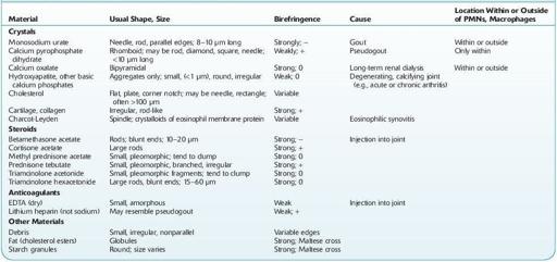
Several types of crystals have been found in synovial fluids (Table 16.26). The two most important are monosodium urate (MSU), characteristic of gouty effusions, and calcium pyrophosphate dihydrate (CPPD), characteristic of the effusions of pseudogout (crystal deposition disease). Other crystals such as calcium hydroxyapatite, calcium oxalate, cholesterol, and corticosteroid esters may also be associated with inflammatory effusions.
Crystals that cause inflammation are usually 0.5 to approximately 20 μm in length, sparingly soluble in water, and capable of being phagocytized. At the peak of inflammation, most are intracellular.

Normal range:
absent (no crystals present).
TABLE 16–26. Birefringent Materials in Synovial Fluid

+, positive birefringence; −, negative birefringence; 0, no axis.
Crystals are best seen in fresh, wet-mount preparations examined with polarizing light.
Hydroxyapatite complexes (diagnostic of apatite disease) and basic calcium phosphate complexes can be identified only by EM; most cases are suspected clinically but never confirmed.
EDTA, ethylenediaminetetraacetic acid; PMN, polymorphonuclear neutrophil.
Other books
The Hunger Games Tribute Guide by Seife, Emily
Predator One by Jonathan Maberry
A Trashy Affair by Shurr, Lynn
Spirit [New Crescent 2] (BookStrand Publishing Romance) by Mary Lou George
In Spite of Thunder by John Dickson Carr
TroubleinChaps by Ciana Stone
The Summer the World Ended by Matthew S. Cox
Dreams of Us by St. James, Brooke
Becoming Three by Cameron Dane