Your Inner Fish: A Journey Into the 3.5-Billion-Year History of the Human Body (16 page)
Read Your Inner Fish: A Journey Into the 3.5-Billion-Year History of the Human Body Online
Authors: Neil Shubin

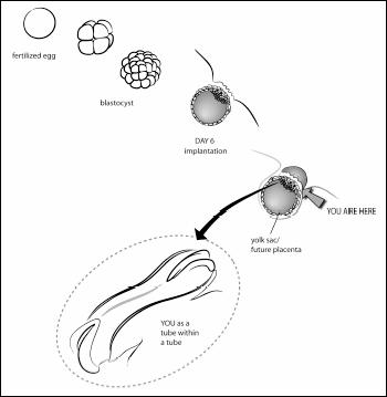
In any event, at this stage of development we are extremely humble-looking creatures. Around the beginning of our second week after conception, the blastocyst has implanted, with one part of the ball embedded in the wall of the uterus, and the other free. Think of a balloon pushed into a wall: this flattened disk becomes the human embryo. Our
entire
body forms from only the top part of this ball, the part that is mushed into the wall. The part of the blastocyst below the disk covers the yolk. At this stage of development, we look like a Frisbee, a simple two-layered disk.
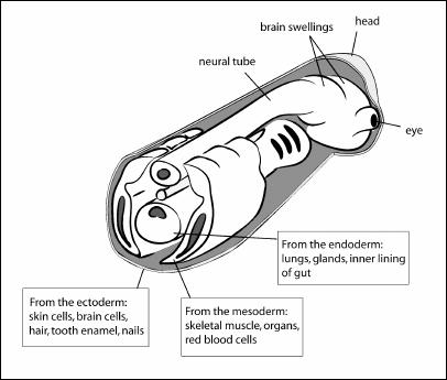
How does this oval Frisbee end up with von Baer’s three germ layers and go on to look anything like a human? First, cells divide and move, causing tissues to fold in on themselves. Eventually, as tissues move and fold, we become a tube with a folded swelling at the head end and another at the tail. If we were to cut ourselves in half right about now, we would find a tube within a tube. The outer tube would be our body wall, the inner tube our eventual digestive tract. A space, the future body cavity, separates the two tubes. This tube-within-a-tube structure stays with us our entire lives. The gut tube gets more complicated, with a big sac for a stomach and long intestinal twists and turns. The outer tube is complicated by hair, skin, ribs, and limbs that push out. But the basic plan persists. We may be more complicated than we were at twenty-one days after conception, but we are still a tube within a tube, and all of our organs derive from one of the three layers of tissue that appeared in our second week after conception.
The names of these three all-important layers are derived from their position: the outer layer is called ectoderm, the inner layer endoderm, and the middle layer mesoderm. Ectodermformsmuch of the outer part of the body (the skin) and the nervous system. Endoderm, the inside layer, forms many of the inner structures of the body, including our digestive tract and numerous glands associated with it. The middle layer, the mesoderm, forms tissue in between the guts and skin, including much of our skeleton and our muscles. Whether the body belongs to a salmon, a chicken, a frog, or a mouse, all of its organs are formed by endoderm, ectoderm, and mesoderm.

Our early days, the first three weeks after conception. We go from being a single cell to a ball of cells and end up as a tube.
Von Baer saw how embryos reveal fundamental patterns of life. He contrasted two kinds of features in development: features shared by every species, and features that vary from species to species. Features such as the tube-within-a-tube arrangement are shared by all animals with a backbone: fish, amphibians, reptiles, birds, and mammals. These common features appear relatively early in development. The features that distinguish us—bigger brains in humans, shells on turtles, feathers on birds—arise relatively later.
Von Baer’s approach is very different from the “ontogeny recapitulates phylogeny” idea you might have learned in school. Von Baer simply compared embryos and noted that the embryos of different species looked more similar to each other than do the adults of those species. The “ontogeny recapitulates phylogeny” approach championed decades later by Ernst Haeckel made the claim that each species tracked its evolutionary history as it proceeded through development. Accordingly, the embryo of a human went through a fish, a reptile, and a mammal stage. Haeckel would compare a human embryo to an adult fish or a lizard. The differences between the ideas of von Baer and Haeckel might seem subtle, but they are not. In the past one hundred years, time and new evidence have treated von Baer much more kindly. In comparing embryos of one species to adults of another, Haeckel was comparing apples to oranges. A more meaningful comparison is one where we can ultimately uncover the mechanisms that drive evolution. For that, we compare embryos of one species to embryos of another. The embryos of different species are not completely identical, but their similarities are profound. All have gill arches, notochords, and look like a tube within a tube at some stage of their development. And, importantly, embryos as distinct as fish and people have Pander and von Baer’s three germ layers.

At four weeks after conception, we are a tube within a tube and have the three germ layers that give rise to all our organs.
All of these comparisons lead us to the real issue at stake. How does the embryo “know” to develop a head at the front end and an anus at the back? What mechanisms drive development and make cells and tissues able to form bodies?
To answer these questions required a whole new approach. Rather than simply comparing embryos as in von Baer’s day, we had to find a new way of analyzing them. The latter part of the nineteenth century ushered in the era, which we first discussed in Chapter 3, when embryos were chopped, grafted, split, and treated with virtually every kind of chemical imaginable. All in the name of science.
EXPERIMENTING WITH EMBRYOS

Biologists at the turn of the twentieth century were grappling with fundamental questions about bodies. Where in the embryo does the information to build them lie? Is this information contained in every cell or in patches of cells? And what form does this information take—is it a special kind of chemical?
Beginning in 1903, the German embryologist Hans Spemann began to investigate how cells learned to build bodies during development. His goal was to find where the body-building information resides. The big question for Spemann was whether all the cells in the embryo have enough information to build whole bodies, or whether that information is confined to certain parts of the developing embryo.
Working with newt eggs, which were easy to obtain and relatively easy to fiddle with in the lab, Spemann devised a clever experiment. He cut off a strand of his infant daughter’s hair and made a miniature lasso out of it. Baby hair is remarkable stuff; soft, thin, and pliant, it made the ideal material for tying up a tiny sphere such as a newt egg. Spemann did exactly that to a developing newt egg, pinching one side off from the other. Manipulating the nuclei of the cells a bit, he let the resulting contraption develop and watched what happened. The embryo formed twins: two complete salamanders emerged, each with a normal body plan and each entirely viable. The conclusion was obvious: from one egg can come more than one individual. This is what identical twins are. Biologically, Spemann had demonstrated that in the early embryo some cells have the capacity to form a whole new individual on their own.
This experiment was only the beginning of a whole new phase of discovery.
In the 1920s Hilde Mangold, a graduate student in Spemann’s laboratory, started to work with small embryos. The fine control she had of her fingers made her able to do some incredibly demanding experiments. At the stage of development with which Mangold worked, the salamander embryo is a sphere about a sixteenth of an inch in diameter. She lopped off a tiny piece of tissue, smaller than a pinhead, from one part of the embryo and grafted it onto the embryo of another species. What Mangold transplanted wasn’t just any patch, but an area where cells that were to form much of the three germ layers were moving and folding. Mangold was so skilled that the grafted embryos actually continued to develop, giving her a pleasant surprise. The grafted patch led to the formation of a whole new body, including a spinal cord, back, belly, even a head.

Just by moving a small patch of tissue in the embryo, Mangold produced twins.
Why is all this important? Mangold had discovered a small patch of tissue that was able to direct other cells to form an entire body plan. The tiny, incredibly important patch of tissue containing all this information was to be known as the Organizer.
Mangold’s dissertation work was ultimately to win the Nobel Prize, but not for her. Hilde Mangold died tragically (the gasoline stove in her kitchen caught fire) before her thesis could even be published. Spemann won the Nobel Prize in Medicine in 1935, and the award cites “his discovery of the Organizer and its effect in embryonic development.”
Today, many scientists consider Mangold’s work to be the single most important experiment in the history of embryology.
At roughly the same time that Mangold was doing experiments in Spemann’s lab, W. Vogt (also in Germany) was designing clever techniques to label cells, or batches of them, and thus allow the experimenter to watch what happens as the egg develops. Vogt was able to produce a map of the embryo that shows where every organ originates in the egg. We see the antecedents of the body plan in the cell fates of the early embryo.
From the early embryologists, people like von Baer, Pander, Mangold, and Spemann, we have learned that all the parts of our adult bodies can be mapped to individual batches of cells in the simple three-layered Frisbee, and the general structure of the body is initiated by the Organizer region discovered by Mangold and Spemann.
Cut, slice, and dice, and you’ll find that all mammals, birds, amphibians, and fish have Organizers. You can even sometimes swap one species’ Organizer for another. Take the Organizer region from a chicken and graft it to a salamander embryo: you get a twinned salamander.
But just what is an Organizer? What inside it tells cells how to build bodies? DNA, of course. And it is in this DNA that we will find the inner recipe that we share with the rest of animal life.
OF FLIES AND MEN
