Breast Imaging: A Core Review (27 page)
Read Breast Imaging: A Core Review Online
Authors: Biren A. Shah,Sabala Mandava
Tags: #Medical, #Radiology; Radiotherapy & Nuclear Medicine, #Radiology & Nuclear Medicine

B. Surgical consultation for duct excision
C. Breast MRI
D. Spot compression–magnification views of the retracted nipple
121
Which of the following types of calcifications can have a radiolucent center?
A. Milk of calcium
B. Ductal carcinoma in situ (DCIS)
C. Calcifications of fibrocystic change
D. Dermal calcifications
122
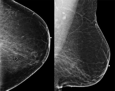
Regarding the finding seen on the image below, which of the following statements is correct? The patient was recently in a car accident.

A. The trauma that causes this injury can be blunt or penetrating.
B. Most common cause of fat necrosis is surgery.
C. Usually is seen in the upper inner quadrant on the right when the trauma is sustained by the driver.
D. History does not help in diagnosis.
123
Which of the following statements concerning neoadjuvant chemotherapy (NAT) for breast cancer is correct?
A. Major use is in primary inoperable locally advanced breast cancer (LABC).
B. Primary goal is to shrink tumor for the potential for breast conservation surgery.
C. Size of tumor is most accurately determined by ultrasound.
D. Accuracy in preoperative size on mammography is best for invasive lobular carcinoma.
124
Which of the following pathologies from core needle biopsy of a lesion, that has been increasing in size, is a reason for surgical excision?
A. Pseudoangiomatous stromal hyperplasia (PASH)
B. Apocrine cyst
C. Fat necrosis
D. Steatocystoma multiplex
125a
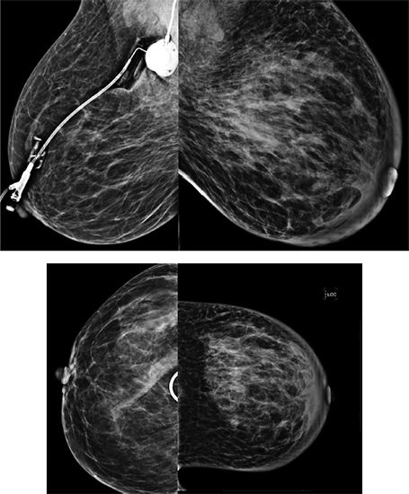
Based on the images below, which of the following statements regarding the findings is correct?

A. Architectural distortion with associated skin thickening
B. Focal asymmetry with associated skin thickening
C. Trabecular thickening with associated skin thickening
D. Segmental calcifications with associated skin thickening
125b
Based on the findings, which of the following diagnoses is the most likely?
A. Invasive ductal carcinoma
B. Reduction mammoplasty
C. Seat-belt injury/trauma
D. Mastitis
126a
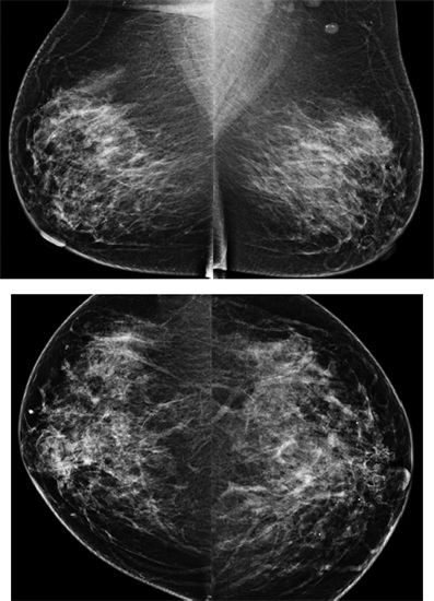
A 40-year-old female presents for baseline screening mammogram.

What is the BI-RADS category?
A. BI-RADS 0
B. BI-RADS 1
C. BI-RADS 2
D. BI-RADS 3
E. BI-RADS 4
126b
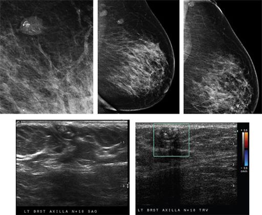
Additional mammographic views and ultrasound images of the mass are shown. Which of the following statements concerning the mass depicted is correct?

A. Core needle biopsy or fine needle aspiration can be performed for diagnosis.
B. Occurs with the same frequency in males and females
C. Up to 5% may undergo malignant transformation.
D. Most common location is in the breast.
127
Which of the following conditions can lead to skin thickening in the breast?
A. Ovarian carcinoma
B. Silicone implant rupture
C. Psoriasis
D. Tuberculosis
ANSWERS AND EXPLANATIONS
1
Answer C.
Intracapsular rupture of a double-lumen breast implant is the disruption or tear of an implant shell in which silicone gel moves outside of the implant shell but stays within the fibrous capsule. Intracapsular rupture is more commonly seen than extracapsular rupture.
Reference: Shah BA, Fundaro GM, Mandava S.
Breast Imaging Review: A Quick Guide to Essential Diagnoses
. 1st ed. New York, NY: Springer; 2010:205–206, 236.
2
Answer A.
The most common location for an intramammary lymph node is in the upper outer quadrant. Approximately 90% of the intramammary lymph nodes are present here.
Reference: Berg A, Birdwell R, Gombos E.
Diagnostic Imaging Breast
. 1st ed. Salt Lake City, UT: Amirsys; 2008;IV:1–8.
3a
Answer C.
The dominant finding is unilateral right breast skin thickening most pronounced medial and lateral to the nipple.
3b
Answer B.
Differential diagnosis for skin thickening includes unilateral edema (focal or diffuse), mastitis, inflammatory carcinoma, postprocedural skin thickening, abscess, and underlying malignancy. Breast parenchymal enhancement may vary with the phase of the menstrual cycle, but skin thickening will not occur.
Reference: Berg A, Birdwell R, Gombos E.
Diagnostic Imaging Breast
. 1st ed. Salt Lake City, UT: Amirsys; 2008;IV:3-26–IV:3-27.
4a
Answer C.
A targeted ultrasound exam is recommended as an initial test for women <30 years of age, pregnant, or lactating.
Reference: Berg A, Birdwell R, Gombos E.
Diagnostic Imaging Breast
. 1st ed. Salt Lake City, UT: Amirsys; 2008;II:0–32.
4b
Answer B.
Major types of fibroadenomas are adult type and juvenile. Most fibroadenomas in teenagers are of the adult type. Giant fibroadenomas are more common in African American women. Giant fibroadenomas are defined as being greater than 8 cm or larger in size. Fibroadenomas are the most common breast mass in women under 35 years of age. They comprise 10% of breast masses in postmenopausal women. They are present almost exclusively in females.
4c
Answer D.
Color Doppler ultrasound demonstrates a solid, hypoechoic, oval, circumscribed, vascular mass with long axis parallel to the skin surface that has an appearance of a fibroadenoma. The most common solid benign tumor in young female is a fibroadenoma. Juvenile fibroadenoma occurs usually been in the age of 10-20, rare above the age of 45. Because juvenille fibroadenomas can grow to a large size, they can be called a giant fibroadenomas. However, not all giant fibroadenomas are juvenile fibroadenomas. The appearance of fat necrosis on ultrasound evolves over time. The sonographic spectrum can range from anechoic, echogenic, irregular hypoechoic mass and a complex cystic and solid mass. Lymph nodes will have an echogenic central vascular hilum on sonography.
References: Berg A, Birdwell R, Gombos E.
Diagnostic Imaging Breast
. 1st ed. Salt Lake City, UT: Amirsys; 2008;IV:2-24–IV:2-35.
Ikeda DM.
Breast Imaging: The Requisites
. 2nd ed. St. Louis, MO: Elsevier Mosby; 2011:117.
5
Answer D.
Unilateral, spontaneous, serous, or bloody nipple discharge is a worrisome clinical finding and warrants imaging evaluation. Intraductal papillomas are epithelial proliferations of the duct, which usually have a central vascular stalk. They are the resultant cause of ~34% of nipple discharge. Fibrocystic change (25%), ductal ectasia (13%), and carcinoma (10%) are in the differential diagnosis of intraductal lesions leading to nipple discharge but are less common than intraductal papillomas.
Reference: Stavros AT.
Breast Ultrasound
. Philadelphia, PA: Lippincott Williams & Wilkins; 2004:157–160.
6a
Answer A.
The images demonstrate round and smudgy calcifications on the CC view that have a curvilinear appearance on the ML view. These are representative of milk of calcium, which are benign. Milk of calcium is sedimented calcium oxalate calcifications within microcysts and dilated lobules.
6b
Answer A.
Milk of calcium is a benign entity, and therefore no further workup or intervention is necessary.
Reference: Shah BA, Fundaro GM, Mandava S.
Breast Imaging Review: A Quick Guide to Essential Diagnoses
. 1st ed. New York, NY: Springer; 2010:28–29.
Other books
Cook the Books by Jessica Conant-Park, Susan Conant
Patricia Dusenbury - Claire Marshall 01 - A Perfect Victim by Patricia Dusenbury
Dragonlance 08 - Dragons of the Highlord Skies by Margaret Weis, Tracy Hickman
After the Abduction by Sabrina Jeffries
On the Mountain by Peggy Ann Craig
Once They Were Eagles by Frank Walton
The Whizz Pop Chocolate Shop by Kate Saunders
From Venice With Love by Alison Roberts
Yours Again (River City Series) by Burks, Dee
La dama del lago by Andrzej Sapkowski