Read Ross & Wilson Anatomy and Physiology in Health and Illness Online
Authors: Anne Waugh,Allison Grant
Tags: #Medical, #Nursing, #General, #Anatomy
Ross & Wilson Anatomy and Physiology in Health and Illness (192 page)


This section considers the main muscles that move the limbs, as well as the major muscles of the face and neck, back, chest, pelvic floor and abdominal wall.
Muscles of the face and neck (
Fig. 16.58
)

Figure 16.58
The main muscles on the right side of the face, head and neck.
Muscles of the face
Many muscles are involved in changing facial expression and with movement of the lower jaw during chewing and speaking. Only the main muscles are described here. Except where indicated the muscles are present in pairs, one on each side.
Occipitofrontalis (unpaired)
This consists of a posterior muscular part over the occipital bone (
occipitalis
), an anterior part over the frontal bone (
frontalis
) and an extensive flat tendon or aponeurosis that stretches over the dome of the skull and joins the two muscular parts. It raises the eyebrows.
Levator palpebrae superioris
This muscle extends from the posterior part of the orbital cavity to the upper eyelid. It raises the eyelid.
Orbicularis oculi
This muscle surrounds the eye, eyelid and orbital cavity. It closes the eye and when strongly contracted ‘screws up’ the eyes.
Buccinator
This flat muscle of the cheek draws the cheeks in towards the teeth in chewing and in forcible expulsion of air from the mouth (‘the trumpeter’s muscle’).
Orbicularis oris (unpaired)
This muscle surrounds the mouth and blends with the muscles of the cheeks. It closes the lips and, when strongly contracted, shapes the mouth for whistling.
Masseter
This is a broad muscle, extending from the zygomatic arch to the angle of the jaw. In chewing it draws the mandible up to the maxilla, closing the jaw, and exerts considerable pressure on the food.
Temporalis
This muscle covers the squamous part of the temporal bone. It passes behind the zygomatic arch to be inserted into the coronoid process of the mandible. It closes the mouth and assists with chewing.
Pterygoid
This muscle extends from the sphenoid bone to the mandible. It closes the mouth and pulls the lower jaw forward.
Muscles of the neck
There are many muscles in the neck, but only the two largest are considered here.
Sternocleidomastoid
This muscle arises from the manubrium of the sternum and the clavicle and extends upwards to the mastoid process of the temporal bone. It assists in turning the head from side to side. When the muscle on one side contracts it draws the head towards the shoulder. When both contract at the same time they flex the cervical vertebrae or draw the sternum and clavicles upwards when the head is maintained in a fixed position, e.g. in forced respiration.
Trapezius
This muscle covers the shoulder and the back of the neck. The upper attachment is to the occipital protuberance, the medial attachment is to the transverse processes of the cervical and thoracic vertebrae and the lateral attachment is to the clavicle and to the spinous and acromion processes of the scapula. It pulls the head backwards, squares the shoulders and controls the movements of the scapula when the shoulder joint is in use.
Muscles of the trunk
These muscles stabilise the association between the appendicular and axial skeletons at the pectoral girdle, and stabilise and allow movement of the shoulders and upper arms.
Muscles of the back
There are six pairs of large muscles in the back, in addition to those forming the posterior abdominal wall (
Figs 16.59
,
16.60
and
16.61
). The arrangement of these muscles is the same on each side of the vertebral column. They are:
•
trapezius (see above)
•
latissimus dorsi
•
teres major
•
psoas (
p. 418
)
•
quadratus lumborum
•
sacrospinalis.

Figure 16.59
The main muscles of the back.

Figure 16.60
The deep muscles of the posterior abdominal wall.

Figure 16.61
Transverse section of the posterior abdominal wall:
a lumbar vertebra and its associated muscles.
Latissimus dorsi
This arises from the posterior part of the iliac crest and the spinous processes of the lumbar and lower thoracic vertebrae. It passes upwards across the back then under the arm to be inserted into the bicipital groove of the humerus. It adducts, medially rotates and extends the arm.
Teres major
This originates from the inferior angle of the scapula and is inserted into the humerus just below the shoulder joint. It extends, adducts and medially rotates the arm.
Quadratus lumborum
This muscle originates from the iliac crest, then it passes upwards, parallel and close to the vertebral column and it is inserted into the 12th rib (
Fig. 16.60
). Together the two muscles fix the lower rib during respiration and cause extension of the vertebral column (bending backwards). If one muscle contracts it causes lateral flexion of the lumbar region of the vertebral column.
Sacrospinalis (erector spinae)
This is a group of muscles lying between the spinous and transverse processes of the vertebrae (
Fig. 16.61
). They originate from the sacrum and are finally inserted into the occipital bone. Their contraction causes extension of the vertebral column.
Muscles of the abdominal wall
Five pairs of muscles form the abdominal wall (
Figs 16.62
and
16.63
). From the surface inwards they are:
•
rectus abdominis
•
external oblique
•
internal oblique
•
transversus abdominis
•
quadratus lumborum (see above).
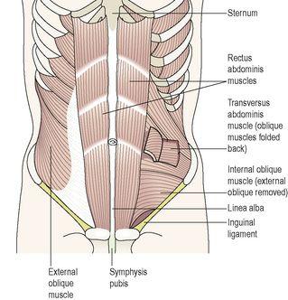
Figure 16.62
The muscles of the anterior abdominal wall.
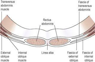
Figure 16.63
Transverse section of the muscles and fasciae of the anterior abdominal wall.
The anterior abdominal wall is divided longitudinally by a very strong midline tendinous cord, the
linea alba
(meaning ‘white cord’) which extends from the xiphoid process of the sternum to the symphysis pubis. The structure of the abdominal wall on each side of the linea alba is identical.
Rectus abdominis
This is the most superficial muscle. It is broad and flat, originating from the transverse part of the pubic bone then passing upwards to be inserted into the lower ribs and the xiphoid process of the sternum. Medially the two muscles are attached to the linea alba.