Breast Imaging: A Core Review (5 page)
Read Breast Imaging: A Core Review Online
Authors: Biren A. Shah,Sabala Mandava
Tags: #Medical, #Radiology; Radiotherapy & Nuclear Medicine, #Radiology & Nuclear Medicine

A. Two fibers, two microcalcification clusters, and two masses
B. One fiber, two microcalcification clusters, and one mass
C. Three fibers, three calcification clusters, and three masses
D. Four fibers, three calcification clusters, and three masses
40
The view shown in the image below is suboptimal for evaluating which portion of the breast?
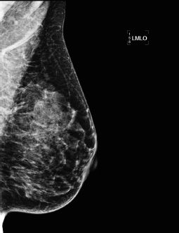
A. Inferior
B. Lateral
C. Medial
D. Superior
41
Regarding contrast-enhanced breast MRI for the detection of breast cancer, which one of the following statements is correct?
A. Cancer is excluded if a mass has hyperintense/fluid signal on the T2-weighted sequence.
B. Breast MRI is optimally performed in week 4 of a patient’s menstrual cycle.
C. T1-weighted non–fat saturation is the best sequence for identification of a fat-containing mass.
D. A body coil is the optimal radiofrequency receiver coil for the exam.
E. An equivalent dose of a gadolinium-based contrast agent is used for breast MR patients.
42a
The following image from a contrast-enhanced breast MR examination demonstrates which artifact?
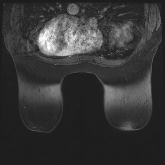
A. Chemical shift artifact
B. Wrap/aliasing artifact
C. Susceptibility artifact
D. Patient motion/ghosting artifact
E. Inhomogeneous fat saturation artifact
42b
What can reduce inhomogeneous fat saturation artifact on breast MRI?
A. Enlarging the field of view
B. Reducing patient motion
C. Shimming the magnet frequently
D. Increasing the bandwidth
E. Check for a leak in the radiofrequency (RF) shield
43
The following image from a contrast-enhanced breast MR examination demonstrates which artifact?
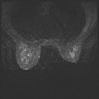
A. Chemical shift artifact
B. Wrap/aliasing artifact
C. Susceptibility artifact
D. Patient motion/ghosting artifact
E. Inhomogeneous fat saturation artifact
44
Which one of the following artifacts is present on the axial postcontrast T1-weighted fat-saturated MR image seen below?
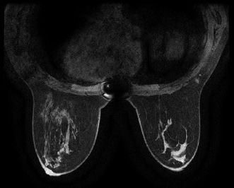
A. Chemical shift artifact
B. Wrap/aliasing artifact
C. Susceptibility artifact
D. Patient motion/ghosting artifact
E. Inhomogeneous fat saturation artifact
45
Based on the images, which one of the following breast imaging ultrasound lexicon terminologies best describes the finding?
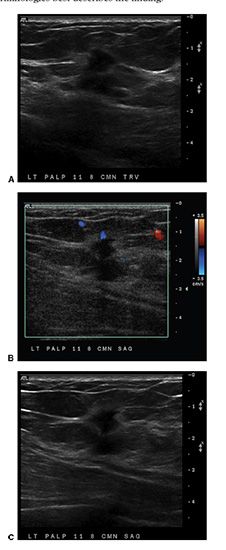
A. Oval isoechoic mass with a circumscribed margin
B. Lobular hypoechoic mass with associated skin thickening
C. Round, anechoic mass with posterior acoustic enhancement
D. Irregular hypoechoic mass with angular margins
46
You are shown a left mediolateral oblique (MLO) and craniocaudal (CC) (zoomed) mammogram images (Figures A and B). What is the MOST descriptive of the calcifications?
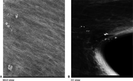
A. Amorphous
B. Pleomorphic
C. Punctate
D. Lucent centered
E. Dystrophic
47
In a well-positioned mammogram, which of the following statements is correct?
A. The pectoralis muscle should be convex on the mediolateral oblique (MLO) view.
B. The pectoralis muscle should extend inferior to the posterior nipple line on the MLO view.
C. The pectoralis muscle thickness should be >1 cm on the craniocaudal (CC) view.
D. The CC view should be exaggerated to include the axillary tail.
E. The length of the posterior nipple line on the CC view should be 1 cm greater than on the MLO view.
48
The mediolateral oblique (MLO) image taken during a screening mammogram examination demonstrates which type of digital mammogram artifact?
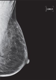
A. Motion artifact
B. Ghost image
C. Grid lines
D. Deodorant artifact
49
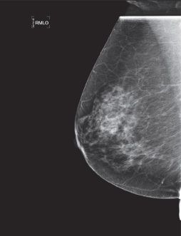
The below mediolateral oblique (MLO) image was taken during a screening mammogram examination demonstrates which type of digital mammogram artifact?
Other books
Sinful Pleasure (The Millionaires Social Club) by VonSant, Kat
Joe and Marilyn: Legends in Love by C. David Heymann
Larry and the Meaning of Life by Janet Tashjian
Sookie and The Snow Chicken by Aspinall, Margaret
Forever Bound by Stacey Kennedy
Nowhere to Run by C. J. Box
La niña del arrozal by Jose Luis Olaizola
Fifty Shades of Jamie Dornan by Louise Ford
Master's Flame by Annabel Joseph
Highway 61 by David Housewright