i bc27f85be50b71b1 (78 page)
Read i bc27f85be50b71b1 Online
Authors: Unknown

Distal radius;
Closed reduction with
See Distal radius;
illtra-articul:ar,
external fixation
intra-articular, above
comminuted
ORIF
Bone grafting
Splinr
:t: = with or widlOuf; l',,rWB = non weight bearing; ORIF = open-reduction internal fixation.
Sou rce: Adapted from DL Fernandez, AK Palmer. Fracture of the Distal Radius. In OP
Green, RN Hotchkiss, WL Pederson (eds), Green's Operative Hand Surgery, Vol. I (4th
cd). New York: Churchill Livingstone, 1998;950.

4
Nervous System
Michele P. West
Introduction
The nervous system is linked ro every system of the body and is
responsible for the integration and regulation of homeostasis. It is
also involved in the action, communication, and higher cortical
function of the body. A neurologic insult and its manifestations
therefore have the potential ro affect multiple body systems. To
safely and effectively prevent or improve the neuromuscular, systemic, and functional sequelae of altered neurologic status in the acme care setting, the physical therapist requires an understanding
of the neurologic system and the principles of neuropathology. The
objectives of this chapter are ro provide the following:
1 .
A brief review of the structure and function of the nervous
system
2.
An overview of neurologic evaluation, including the physi-
cal examination and diagnostic tests
3.
A description of common neurologic diseases and disor-
ders, including clinical findings, medical and surgical management,
and physical therapy interventions
259

260 AClffE CARE HANDBOOK FOR PHYSICAL ll-IERAPISTS
Structure and Function of the Nervous System
The nervous system is divided as follows:
1 .
The central nervous system (CNS), consisting of the brain
and spinal cord
2.
The peripheral (voluntary) nervous system, consisting of
efferent and afferent somatic nerves outside the CNS
3.
The autonomic (involuntary) nervous system, consisting of
the sympathetic and parasympathetic systems
Central Neroous System
Brain
The brain is anatomically divided into the cerebral hemispheres,
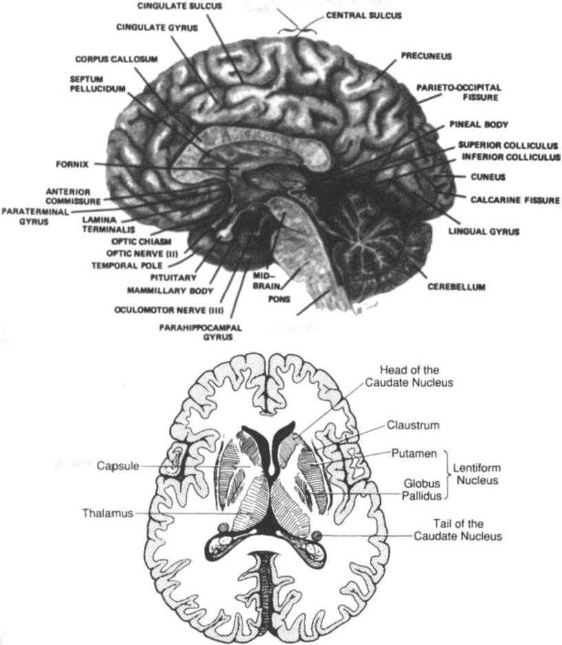
diencephalon, brain stem, and cerebellum. A midsagittal view of the
brain is shown in Figure 4-1A. Although each portion of the brain
has its own function, it is linked to other portions via traces and
rarely works in isolation. When lesions occur, disruption of these
functions can be predicted. Figure 4- 1 B shows the basal ganglia and
rhe internal capsule. Tables 4-1 and 4-2 describe the basic structure,
function, and dysfunction of the cerebral hemispheres, diencephalon, brain stem, and cerebellum.
Protective Mechanisms
The brain is protected by the cranium, meninges, ventricular system,
and blood-brain barrier.
Cranium
The cranium encloses the brain. It is composed of eight cranial and
14 facial bones connected by sutures and contains a pproximately
85 foramen for the passage of the spinal cord, cranial nerves
(CNs), and blood vessels.' The cranium is divided into rhe cranial
vault, or calvaria (the superolateral and posterior aspects), and the
cranial floor, which is composed of fossae (the anterior fossa sup
POrtS the frontal lobes; the middle fossa supports the temporal
lobes; and the posterior fossa supports the cerebellum, pons, and
medulla).2

NERVOUS SYSTEM
261
............... """'.
-""" ..
MlClIO'ML",., I'OITCltmlAL
GnU)
.. .......
A
Inlemal
B
Figure 4-1. A. Medial (midsagittal) view of a hemisected brain. (\Vith permission from S Gilman, S W Newman ledsJ. Manter and Gatz's Essentials of Neuroanatomy and Neurophysiology [7th edJ. New York: Oxford University
Press, 1987;9.) B. Horizontal section of the cerebrum showing the basal gang[ia. ( With permission from RJ Love. we Webb [eds[. Neuro[ogy for the Speech-Language Pathologist [4th ed]. Boston: Butte nvorth- Heinemaml,
2001 ;38.)


Table 4-1. Structure, Function, and Dysfunction of the Cerebral Hemispheres
N
'"
N
Lobe of
>
Cerebrum
Structure
Function
Dysfunction
g '"
Frontal
Precentral gyrus
Voluntary mmor cortex of contralateral
Contralateral mono- or hemiparesis
()
>
lobe
face, arm, trunk, and leg
or hemiplegia
"
'"
Supplementary
Advanced motor planning
Conualateral head and eye paralysis
J:
>
motor area
Contralateral head and eye turning
Z
"
'"
(connections to cranial nerves Ill, N, VI,
"
"
IX, X, and Xll nuclei)
'"
�
Prefrontal pole
Personaliry center, including abstract ideas,
Loss of inhibition and demonstradon of
"
"
concern for mhers, conscience, initia-
antisocial behaviors
�
J:
rive, judgment, persistence, and planning Ataxia, primitive reflexes, and hypertoniciry
-<
�
Paracentral
Bladder and bowel inhibition
Urinary and bowel incontinence
n
>
lobule
r
-l
Broca's a rea
D: Mmor speech center
Broca's (expressive) aphasia
J:
'"
"
ND: Appreciation of intonation and
>
�
gestures with vocalization
�
Parietal
Postcentral gyrus
Somatosensory cortex of contralateral
Contralateral sensation loss
lobe
pain, posture, proprioception, and touch
of arm, trunk, and leg
Parietal pole
D: Abiliry to perform calculations
D: Acalculia, agraphia, finger agnosia