Secondary Schizophrenia (4 page)
Read Secondary Schizophrenia Online
Authors: Perminder S. Sachdev

mately 60% of the studies in schizophrenia have shown
Edinburgh
[16],
New York
[17],
Copenhagen
[18],
and
hypo-activation in the prefrontal cortex during various
Israel
[19]).
The New York High-Risk Study demon-working memory tasks
[27].
However, abnormalities
strated that deficits in attention, motor skills, and
of prefrontal cortical function in schizophrenia are not
short-term memory detected between the ages of 7
reducible to simply too much or too little activity and
and 12 years predicted development of schizophrenia-may reflect a compromised effort in processing infor-related psychosis in 58%, 75%, and 83% of cases,
mation mediated by the DLPFC
[28].
respectively
[20].
Similarly, verbal memory and execu-Some of the electrophysiological abnormalities in
tive function predicted later development of psychosis
patients with schizophrenia and their relatives include
in the Edinburgh High Risk Study
[21].
Together, these
abnormal smooth-pursuit eye movements, persistent
observations suggest that cognitive impairments pre-deficit in performance on the antisaccade tasks
[29],
cede the emergence of typical symptoms by several
abnormalities in the auditory evoked potential (i.e.
years.
diminished amplitude and increased latency in the
P300 response to an “oddball” auditory stimulus),
How do neurocognitive alterations
prepulse inhibition of the startle reflex (which measures the ability of a preceding weak prestimulus to
originate? Studies of brain function
transiently inhibit the response to a closely following
Alterations in brain function in schizophrenia have
strong sensory stimulus), and reduced high frequency
been documented on functional neuroimaging stud-
(in the gamma range, i.e., 30–70 Hz) oscillatory power
ies, such as Positron Emission Tomography (PET)
in response to auditory stimulation
[30].
and Blood Oxygenation Level Dependent (BOLD)-
In recent years, the role of the anterior cingu-based functional MRI (fMRI) performed while the
late cortex in cognitive control and self-monitoring
subject is engaged in cognitive tasks. In approximately
has been well recognized
[31]
. Using a test of selec-half of patients with schizophrenia, decreased frontal
tive attention (i.e. the Stroop task), patients with
metabolism and blood flow are evident during cogni-schizophrenia showed reduced error monitoring and
tive activation tasks. N-back task, which involves the
reduced anterior cingulate activity compared to con-subject observing a sequence of letters and respond-trols
[32, 33].
Earlier, Fletcher and colleagues had
ing to a reappearance of a letter after n trials (i.e., n
=
shown disruption of the normal anterior cingu-0, 1, 2, and so on) is one of the most commonly
late modulation of prefrontotemporal integration in
used tasks. In general, functional neuroimaging stud-patients with schizophrenia
[34].
ies have demonstrated reduced activation of lateral
The amygdala and hippocampus seem to have
prefrontal regions during the task performance
[22].
complementary roles in cognitive processing with the
Recently, a meta-analysis indicated that even subjects
former regulating emotion and affect and the latter,
at genetic risk for schizophrenia show abnormalities
episodic and associative memory
[35, 36].
Subjects
on functional neuroimaging studies
[23].
An fMRI
with amygdala damage have been shown to have an
study has also noted that auditory hallucinations are
impaired ability to interpret facial expressions in a
5
associated with activation in many brain areas, such as
pattern similar to what was shown earlier in autism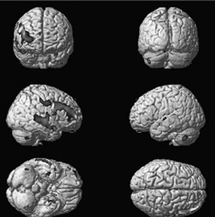
Introduction – Section 1
[37].
Most fMRI studies of amygdala function indicate
that dysfunction of the emotional aspect of the brain
is the hallmark of schizophrenia
[38].
For instance,
patients performed poorly on affect labeling tasks
[39]
and displayed reduced responsivity of the amygdala
[6,
40]
. The relationship between these deficits and
social functioning and the trajectory of these deficits in
at-risk populations are prime areas for further investigation. Taken together, functional imaging studies
point to abnormalities in prefrontal, cingulate, and
medial temporal lobe function early in the illness.
Much work remains to be done to understand the full
implications of each of these observations.
In which part of the brain can abnomalities
be found? Structural brain imaging studies
The early manifestations of cognitive neurofunctional
Figure 1.1
Brain MRI images showing grey-matter volume
deficits, as previously outlined, strongly point to neu-reductions in first-episode schizophrenia subjects compared to
roanatomical alterations in patients with schizophre-healthy controls using Voxel Based Morphometry (VBM). [From the
nia. In vivo neuroimaging studies demonstrate a num-University of Pittsburgh.] (See color plate section.)
ber of brain structural abnormalities in schizophrenia.
Systematic reviews and meta-analyses of MRI stud-Studies of relatives of patients with schizophrenia
ies in schizophrenia indicate reductions in volume
provide further insight into the illness. ROI studies
of whole brain, as well as grey matter volumes and
of offspring and siblings of patients show volumet-increases in ventricular volume
[41, 42, 43, 44].
More
ric reductions in amygdala and hippocampus
[55, 56,
prominent reductions are seen in temporal lobe struc-
57].
Similarly, computational VBM studies have shown
tures, especially in the hippocampus, amygdala, and
reduced grey matter in the PFC in that population
[58,
the superior temporal gyri
[45, 46]
, the prefrontal cor-
59]
. A longitudinal VBM study demonstrated that a
tex, and the thalamus
[47].
Automated regional par-spatial pattern of reductions in grey matter density in
cellation and voxel-based morphometry (VBM) tech-the left temporal lobe and right cerebellum could pre-niques have largely validated this region of interest
dict onset of psychotic symptoms in those at genetic
(ROI)-based findings. Reductions in medial tempo-risk
[60].
ral lobes and the superior temporal gyrus (STG) are
Structural changes have also been shown to pre-well-replicated findings in VBM studies
[48].
More-dict development of psychosis in those with prodromal
over, STG volumes and reductions in medial temporal
symptoms. For instance, Pantelis and colleagues
[61]
volumes correlate with positive symptoms and mem-showed that lesser grey matter in the right medial tem-ory impairment, respectively
[49, 50].
poral, lateral temporal, inferior frontal cortex, and the
Studies of first-episode schizophrenia show that
cingulate cortex bilaterally predicted the one-third of
brain structural alterations are present at illness onset
individuals with prodromal symptoms who developed
[51]
(Figure 1.1).
Two recent meta-analyses of such
psychosis on follow-up. This MRI follow-up study sug-studies
[41]
show whole brain and hippocampal vol-gests an ongoing disease process during the transi-ume reductions. Brain structural changes evident at ill-tion from prodrome to psychosis. Prospective MRIs
ness onset appear to persist during the course of the
in childhood-onset schizophrenia
[62, 63]
reveal a rel-schizophrenic illness. Some
[52, 53]
, but not all
[54],
atively more rapid loss in superior frontal and tem-found evidence for further progression of the struc-poral cortices (
∼
3–4% loss per year as opposed to a
tural deviations. Collectively, imaging studies suggest
more subtle 1–2% decrease per year in matched con-that brain structural alterations are a persistent trait of
trols). A recent review of the literature suggests that
6
schizophrenia.
all of the reduction may not be accounted for by the
Chapter 1 – Neurobiology and etiology of primary schizophrenia: current status
neurons but could be related to white matter changes,
psychoses. A small number of studies of PET scans
that is, demyelination and changes in the lipid
in first-episode schizophrenia have produced sugges-metabolism
[64].
Many studies of childhood-onset
tive evidence of increased dopamine turnover
[71].
schizophrenia support the post-illness onset progres-An early review of 17 PET and postmortem studies
sion model.
revealed a substantive effect size (1.47) for increases
A follow-up study of the nonpsychotic siblings
in D2 receptor density and affinity in schizophrenia
of patients with childhood-onset schizophrenia has
[72].
Presynaptic DA turnover, as measured by striatal
yielded thought-provoking results
[65].
It was not
Fluoro-DOPA uptake, also appears to be increased in
entirely surprising to find grey matter deficits in right
schizophrenia, especially during psychotic exacerba-prefrontal and inferior parietal cortices or even greater
tions
[73].
These findings provide support to the long-reductions in the left prefrontal and bilateral tempo-held view that psychosis may be related to dopaminer-ral cortices. But it was striking to note that these cor-gic hyperfunction in mesolimbic brain regions
[74].
tical deficits in siblings disappeared by age 20 and that
The hyperdopaminergic model of schizophrenia,
attenuation of deficits over time correlated with overall
however, does not explain the cognitive impairments
functioning at the last scan. Thus, early prefrontal and
and the negative symptoms that characterize this ill-temporal grey matter loss appears to be a trait marker
ness. Weinberger
[75]
suggested that schizophrenia
with differential subsequent trajectory of development
may be characterized by a deficit in the mesocortical
among siblings and between those who do and do not
dopaminergic system, which leads to a disinhibition of
develop the disease.
the mesolimbic dopaminergic system, accounting for
As outlined above, structural abnormalities in
positive psychotic symptoms.
schizophrenia are evident in multiple interconnected
Magnetic resonance spectroscopy (MRS) has
brain regions, perhaps suggesting disrupted connec-emerged as an important noninvasive tool to longi-tivity. Diffusion tensor imaging (DTI)
[66],
a reliable
tudinally evaluate neurochemical changes in schizo-method of studying brain connectivity and white mat-phrenia. The majority of MRS studies in schizophre-ter integrity, measures the orientation of water diffu-nia have employed 1H (Proton) MRS. A recent
sion along the axis of tissue elements, such as axons,
meta-analysis and systematic review of in vivo 1H
and has provided further evidence of parallels between
spectroscopy studies in schizophrenia shows reduced
regional development of prefrontal connectivity and
N acetyl aspartate (NAA), a marker of neuronal
cognitive development. DTI studies suggest that work-integrity, primarily in the PFC and hippocampus both
ing memory capacity and performance on cognitive
in first-episode and chronic schizophrenia patients
control tasks correlate with prefrontal
−
parietal con-
[76]
. However, there are negative studies as well
nectivity
[67]
and frontostriatal connectivity, respec-