Welcome to Your Brain (5 page)
Read Welcome to Your Brain Online
Authors: Sam Wang,Sandra Aamodt
Tags: #Neurophysiology-Popular works., #Brain-Popular works

brain can generate dreams, memory, breathing, and every mental process in your life may seem hard
to believe—but it’s true.
This is particularly impressive in view of the brain’s size. Considering its many functions, the
brain is packed into a very small space. Billions of neurons and additional supporting cells
communicate with one another using an astronomical number of synaptic connections—and the entire
operation fits into an object weighing about three pounds, the size of a small cantaloupe.
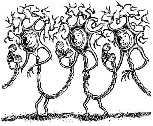
Like a cantaloupe—and the rest of your body—your brain is made of cells. Brain cells come in
two types: neurons, which talk to one another and to the rest of the body, and glial cells, which
provide essential support to keep the whole show going. Your brain is made up of about one hundred
billion neurons—which have a long, skinny, complicated shape—and many more glial cells. From a
distance, the brains of different animals do not look alike. (Compare the shrew and whale brains in
the picture.) They all work according to the same principles, however.
Signals within a neuron are carried by electricity. Each neuron has a net excess density of
negative charge on the inside of the membrane that surrounds it relative to the outside, due to an
uneven distribution of positive and negative ions like potassium and chloride. This unequal
distribution of charge creates a voltage difference across the membrane, like a much smaller version
of the voltage difference that allows a nine-volt battery to give a shock to your tongue. (Actively
moving ions across the membrane to maintain this charge distribution requires more energy than
anything else that the brain does.)
To send electrical signals from one part of the neuron to another, the neuron opens channels that
allow the ions to move across the membrane, creating a current that carries an electrical signal down
the membrane. Neurons receive inputs through branched, treelike structures called dendrites, which
put together information from a bunch of different sources. The neuron then sends an electrical signal
down a long, wirelike structure, called an axon, which triggers a chemical signal to another neuron,
and so on. Axons can carry signals over long distances; your longest axons run from your spine to the
tips of your toes. In contrast, the longest known axons in whales are sixty feet (about twenty meters) in
length. The longest axons belonging to the shrew, whose brain is pictured on the penny, are a mere
two inches (about five centimeters). In all cases, electrical signals spread using similar molecules
and according to the same biological principles.
Did you know? Your brain uses less power than your refrigerator light
Neurons and synapses are so efficient that the brain uses only twelve watts of power—
yet it can do a lot more than the little light in the back of your refrigerator. Over the course
of a day, your brain uses the amount of energy contained in two large bananas. Curiously,
even though the brain is very efficient compared to mechanical systems, in biological terms,
it’s an energy hog. The brain is only 3 percent of the body’s weight, but it consumes one-
sixth (17 percent) of the body’s total energy. Unfortunately, that doesn’t mean that you
should snack more to keep your energy up when you’re studying. Most of the brain’s energy
costs go into maintenance, keeping you ready to think by maintaining the electric field
across each neuron’s membrane that allows it to communicate with other neurons. The
added cost of thinking hard is barely noticeable. Look at it this way: you’re always paying
to support your brain, so you might as well use it!
Let’s look at this process in more detail. Neurons pass information down their axons by
generating small electrical signals that last a thousandth of a second. These signals are called
“spikes” because they represent sudden increases in the electrical currents in a neuron (see graph).
Spikes—known to brain geeks as action potentials—look the same whether they come from squid,
rats, or Uncle Fred, making them a huge success story in the evolutionary history of animals. Racing
down axons at speeds up to several hundred feet per second, spikes bring signals from your brain to
your hand fast enough to escape the bite of a dog or the heat of a frying pan. They help all animals get
away from imminent danger—fast.

Spikes conclude their business when they arrive at the axon’s end. At that point, neurons assume
their other identity, as chemical signaling machines. Each neuron in the brain receives chemical
signals from some neurons and sends chemical signals to others. Communication between neurons
relies on chemicals called neurotransmitters, which are released from small areas at the end of the
axon when triggered by the arrival of a spike. Every neuron makes and receives up to several hundred
thousand chemical connections, called synapses, with other neurons. Neurotransmitters stick to
synaptic receptors on the dendrites or cell bodies of another neuron, triggering further electrical and
chemical signals. All these steps, from release to detection, can take place in a thousandth of a
second.
Synapses are the essential components of communication in your brain. Your thought patterns,
basic abilities and functions, and individuality are determined by how strong these synapses are, how
many of them you have, and where they are. Just as connections in computers mostly connect internal
components of the computer with one another, neurons mostly use synapses to talk to each other
within the brain. Only a small fraction of axons form their synapses outside the brain or spinal cord,
sending signals to other organs of the body, including the muscles.
In addition to being fast, synapses are also very small. The dendritic tree of a typical neuron is
about two-tenths of a millimeter wide. Yet it receives up to two hundred thousand synaptic inputs
from other neurons. Indeed, a cubic millimeter of your brain contains as many as a billion synapses.
Individual synapses are so small that they contain barely enough machinery to function and are
unreliable, so that arriving spikes often fail to cause any release of neurotransmitter at all.
Did you know? Loewi’s dream of the neurotransmitter
Back in 1921, it wasn’t clear how neurons, or even cells in general, talked with one
another. German scientist Otto Loewi made a key observation when he studied how the
heart receives signals to speed up or slow down. He was convinced that the vagus nerve, a
long nerve that comes from the brainstem and attaches to the heart, secreted a substance to
slow the heartbeat. In his laboratory, he carefully dissected the hearts of frogs with the
vagus nerve attached. When he stimulated the vagus nerve with electric shocks, the heart
slowed down. How did this happen? Loewi’s hypothesis was that something came out of
the nerve to cause this effect, but he didn’t know how to test this idea with an experiment.
Stuck, he did what many people do: he slept on it. One night he woke up, struck with an
insight on how to do the experiment. Satisfied, he went back to sleep. The next morning …
nothing. He couldn’t recall what experiment to do. The next time he had the dream, he took
care to write down his idea. Unfortunately, the next morning he couldn’t read his own
writing. Luckily, he had the dream again. This time he didn’t wait: he got up, went to the
laboratory, and did the experiment that would win him the Nobel Prize in Physiology or
Medicine in 1936.
The experiment was a simple one. He placed two frog hearts in two vessels joined by a
narrow tube. One heart had the vagus nerve still attached. When he electrically stimulated
the heart with the nerve attached, it slowed down. Then, after some delay, the second heart
began to slow down as well. This simple experiment demonstrated the existence of what he
unpoetically called
Vagusstoff
, a substance (
stoff
) that comes out of the vagus nerve of one
frog heart to slow the beat of the other heart.
Vagusstoff
, now called acetylcholine, is one
of dozens of neurotransmitters that neurons use to communicate with one another.
It’s odd that synapses are small enough to be flaky, but this appears to be a widespread
phenomenon. Synapses reach a similar minimum size in the brains of various animals, including mice
and people. No one is sure why individual synapses have evolved to be small and unreliable, but one
possible reason is that the brain may work better if it’s packed with a tremendous number of them.
This may be a trade-off that stuffs the most function into the smallest possible space.
For the brain to accomplish its many duties, neurons have to take on very specific tasks. Each
neuron responds to a small number of events, such as hearing a particular sound, seeing someone’s
face, carrying out a certain movement—or other processes that aren’t observable from the outside. At
any given moment, only a small fraction of your neurons, distributed all over your brain, are active.
This fraction is ever shifting; the whole game of thinking depends on which neurons are active and
what they are saying to each other and to the world.
Neurons in all animals are organized into local groups that serve the same broad purpose, such as
detecting visual motion or planning eye movements. In our own brains, each division can have
billions of neurons, with many subdivisions; in a rat, millions, with fewer subdivisions; in a squid or
insect, thousands of neurons (though in these tiny creatures’ brains, different parts of individual
neurons may do multiple things at once). Each of these divisions contains its own distinctive types of
neurons, particular patterns of connection, and connections with other brain structures.
Scientists first learned about the functions of different parts of the brain by studying people with
brain damage. Sadly, World War I was an especially rich source of data. Soldiers often survived
head wounds because high-velocity bullets cauterized their wounds, preventing a fatal loss of blood
or even infection. But the soldiers exhibited a baffling range of symptoms, which depended on the
location in the brain that was damaged. Modern neurologists still publish papers on patients who
have brain damage, most commonly from strokes. Indeed, a few patients with very rare types of
damage actually support themselves by participating in paid studies.
Scientists can also figure out what a neuron does by tracking its activity under different
conditions, by stimulating it, or by tracing its connections to other brain areas. For example, motor
neurons in the spinal cord receive signals from neurons in the cortex that generate basic movement
commands. In turn, these spinal cord neurons send signals to the muscles, causing them to contract. If
scientists electrically stimulate only the spinal cord neurons, the same muscles contract. Together,
these results make it clear that spinal cord motor neurons are responsible for executing movement
commands that are generated at higher levels of the brain, although there is still plenty of controversy
over exactly what aspect of the movement is specified by these commands.
To learn to get around in your brain, you need a quick tour of its parts and what they do. The
brainstem
, as the name suggests, is at the very bottom of the brain, where it attaches to your
spinal
cord
. This region controls basic functions that are critical for life, like reflexive movements of the