Welcome to Your Brain (9 page)
Read Welcome to Your Brain Online
Authors: Sam Wang,Sandra Aamodt
Tags: #Neurophysiology-Popular works., #Brain-Popular works

Touching All the Bases: Your Skin’s Senses
Looking Out for Yourself: Vision
While skiing downhill one day, Mike May realized he was headed toward a huge dark object too
close to dodge. He was sure he was going to die. When he passed through the object, he realized it
was a shadow cast by the ski lift.
Such experiences are common in May’s life, ever since he had his sight restored by a corneal
transplant at age forty-three. May had been blind since a jar of lantern fuel exploded in his face when
he was three. However, blindness did not stop him from becoming an excellent skier. He had claimed
the world record for speed as a blind downhill skier, following his guide down the mountain at sixty-
five miles per hour. During his four decades of blindness, though, his brain had no experience of
natural vision. Now, with his vision restored, he has trouble interpreting what he sees. It’s especially
hard for him to distinguish two-dimensional objects from three-dimensional objects, an essential skill
when you are approaching a large two-dimensional shadow.
Your brain interprets many scenes without making you explicitly aware of what’s going on.
Because May learned to see late in life, the way you might learn a foreign language as an adult, his
brain is unable to accomplish many visual tasks correctly, such as figuring out that the large, dark,
featureless object in front of him was probably a shadow and not a rock. In general, it’s hard for him
to figure out which lines or colors are part of one object, and which are part of another object, or
even part of the background behind the objects. His case illustrates how difficult and important these
processes are in understanding how to see—and how many invisible assumptions your brain needs to
make to get the job done.
Did you know? Animal research and “lazy eye”
One of the best examples of how animal studies can have unexpected benefits for human
medicine comes from research on visual development. Because the two eyes are in
different places on the head, they see the world from slightly different angles. This creates a
problem for brain development; to create a coherent view, the brain needs to match up the
information arriving in the two eyes that comes from the same part of the visual world. It
would be hard to specify this matching in advance, since everyone’s head is a different
size, and the distance between the eyes changes as the body grows. So the brain figures it
out by learning to match up information from locations in each eye that are active at the
same time, and so presumably are seeing the same place in the visual world. If an animal is
deprived of sight in one eye when it’s young, then this learning can’t happen, and almost all
the visual neurons in the brain end up carrying signals from just one eye. If an animal loses
sight in one eye at certain young ages (about the first month after birth in cats, longer for
people), its brain will learn to interpret information only from the other eye. This pattern
can’t be reversed later in life. David Hubel and Torsten Wiesel won the Nobel Prize for
discovering this process.
A friend of ours has a daughter with strabismus, what people used to call lazy eye,
which occurs in 5 percent of children. She has trouble controlling the movement of one eye,
leading it to wander off in a different direction from the other one. Twenty years ago, the
standard treatment for this problem would have been to keep a patch over the good eye (to
train the bad eye to see better). Because of these animal studies, which were undertaken for
pure scientific curiosity, we now know that this treatment isn’t a good idea, even though it
seemed sensible enough at the time. Patching one eye damages brain development because
the brain can’t learn how to process information from the two eyes together.
You need information from both eyes to judge distances. If you close one eye, and then
open that one and close the other, you’ll see that the difference between the views is bigger
for objects that are closer, and smaller for objects that are very far away. Children who are
raised with a patched eye can’t compare information from the two eyes, and they have
trouble with depth perception as adults. For example, they find it extremely difficult to
thread a needle. Because of the animal research, our friend’s daughter is being treated with
a new training procedure that will let her learn to control her eye muscles without
interfering with her ability to see the world in three dimensions later in life.
Vision begins in the eye, which is set up like a camera. A lens in the front of the eye focuses light
onto a thin sheet of neurons in the back, called the retina. Retinal neurons are arranged like a sheet of
pixels, each of which detects the intensity of light in a certain region of the visual world. But this
causes a problem for the brain, because the retina transforms the three-dimensional world into a
pattern of activity in a two-dimensional sheet of neurons, throwing away a lot of the information that’s
out there. (You may have heard that the retina turns the world upside down, which is true, but it
doesn’t affect our vision because the brain expects that and interprets the information correctly.)
Three different types of so-called cone cells in the retina detect red, green, or blue colors in
bright light; these neurons send increasingly strong signals as the intensity of the light that they detect
becomes stronger. Other colors are formed by different levels of activity in combinations of these
three cell types. The process is similar to making many colors of paint by mixing primary colors
together, but the primary colors are different because light mixes differently from paint. (To see for
yourself, put red and green plastic over a couple of flashlights and shine them on the same spot to
make yellow light. Mixing red and green paint gives a very different result, brown.) A fourth cell
type, called a rod, detects light intensity in dim light but does not contribute to color vision, which is
why you can’t see colors as well when the lighting is romantic. These rods and cones then
communicate with other neurons in the retina, which make additional calculations about the scene.
For example, the output cells of the retina carry information about each region’s relative brightness
compared to nearby areas, not about the absolute brightness of each pixel. This information is then
sent into visual areas of the brain, as well as into areas that control movements of the eyes and head.
At each step along the way, neurons are arranged into a map of the visual world, so that
information from neighboring points in the scene is represented by the pattern of spikes in neurons
neighboring each other in each visual brain area. This is similar to the way that points that are close
together in a scene are also close together in a photograph of the scene. Such an organization makes it
easier for neurons that represent nearby parts of the visual world to communicate with each other
when they’re trying to understand their local region of the scene.
The brain must begin by determining the brightness of each part of the object that produced the
visual image. You might imagine that this is a simple task—merely a matter of determining how much
activity is generated in the neuron that transmits information from that part of the scene. However, this
is actually very difficult because neural activity depends on the actual amount of light that reaches the
eye, which varies enormously with the characteristics of the object and with the pattern of
illumination and shadows in the scene. The same object looks very different in bright sun than under a
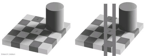
desk lamp, and different again depending on which part of it is in shadow. The figure on the next page
shows that by the time you become aware that you’re seeing an image, the brain has already made a
bunch of assumptions about the object you’re looking at.
In the figure on the left, it’s obvious that the square marked A is darker than the square marked B
—or is it? The figure on the right shows clearly that those two squares have the same shading. Don’t
believe us? Cut a piece of paper to cover the extra squares in the left figure and see for yourself.
Have you ever seen a dog moving its head back and forth while staring at something? A lot of
animals use this trick to figure out the distance of an object. Closer objects appear to move farther
from side to side during this head movement, while more distant objects move less. The brain
calculates depth in a scene from many different cues—and a liberal dose of assumptions. For
example, depth can be calculated by comparing the views from the two eyes or by determining which
objects are in front of other objects. A gravel road going into the distance has two prominent depth
cues: the gravel pieces look smaller when they’re farther away, and the road edges look closer
together. The brain can also use the size of a known object to guess the size of other objects.
Another thing your brain decides automatically is which objects are in a visual image. Mike May
has a lot of trouble identifying objects. He can tell the difference between a triangle and a square
sitting separately on a table, but he has no idea how many people are in a photograph. The skylights at
the mall produce a pattern of alternating bright stripes and shadows across the floor that look, to his
brain, exactly like a staircase. After the operation, his wife had to remind him again and again not to
stare at women, since he can’t get any information from a quick sideways glance the way most men
do. He’s learned intellectually how to reason through a visual scene and figure out what’s in it, to
some extent, but this process will never be fast or effortless for him as it is for most of us.
The brain has special ways of recognizing objects of particular importance to us, such as faces.
The physical differences between faces aren’t all that large—or at least they wouldn’t seem that way
to a Martian—but we can tell them apart effortlessly. People have tried to devise automated face-

recognition systems to identify suspected terrorists in airports and at immigration checkpoints, but
their accuracy is terrible compared with human observers. You can see for yourself that your brain
treats faces in a special way by looking at the pictures of Margaret Thatcher. The photos at the top
look fairly normal to most people—except for being upside down, of course. The bottom pictures are
the same images turned right side up, and now you can see that the one on the right is really weird!
Both the eyes and the mouth have been turned upside down within the face, but you probably didn’t
notice that when looking at the top right picture. Of course, which version you prefer may depend on
other factors, such as your political orientation.
Mike May can’t recognize faces at all. He once offered to buy ice cream for a player after Little
League practice; only when the puzzled boy politely declined his offer did Mike realize that the
player was not his son. Some people who are otherwise normal have the same problem, usually
resulting from damage to a brain region called the fusiform face area, which is responsible for the
specialized processing of faces. These people can see most objects just fine, but they have a lot of
trouble telling people apart, even people they’ve lived with for years. After a while, most of them
learn to memorize what their friends, spouses, or children are wearing when they leave the house so