Examination Medicine: A Guide to Physician Training (98 page)
Read Examination Medicine: A Guide to Physician Training Online
Authors: Nicholas J. Talley,Simon O’connor
Tags: #Medical, #Internal Medicine, #Diagnosis

FIGURE 16.95
Dermatomes of the lower limb.
HINT
Sensory changes can be variable. If sensory loss is not quite consistent but seems generally to fit a pattern, it may be a good idea to describe it confidently as ‘patchy’.
11.
If there is a peripheral sensory loss, attempt to establish a sensory level on the abdomen.
12.
Examine the saddle region sensation (S3–5).
13.
Ask to test the anal reflex (S2–4); if intact, there is brief contraction of the external sphincter of the anus to scratching of the perianal skin.
14.
If you have not done this already go to the back. Look for deformity, scars and neurofibromata. Palpate for tenderness over the vertebral bodies and auscultate for bruits. Test straight leg raising.
15.
It may be relevant to ask whether you can proceed to the upper limbs and cranial nerves.
Notes on the neurological examination of the limbs
Grading muscle power (Medical Research Council)
0.
Complete paralysis.
1.
Flicker of contraction.
2.
Movement with
no
gravity.
3.
Movement with gravity only (any resistance stops movement).
4.
Movement with gravity plus some resistance.
5.
Normal power.
This grading is weighted towards severe weakness (grades 0–3 are all severe). A more sensible scale would be the following:
1.
Complete paralysis.
2.
Severe weakness.
3.
Moderate weakness.
4.
Mild weakness.
5.
Normal.
Signs of a lower motor neurone lesion
1.
Weakness.
2.
Wasting.
3.
Hypotonicity.
4.
Decreased or absent reflexes.
5.
Fasciculation (prominent in anterior horn cell diseases unless far advanced).
Signs of an upper motor neurone lesion
1.
Weakness in an ‘upper motor neurone pattern’; all muscle groups are weak, but may be more marked in upper limb abductor and extensor muscles – shoulder abduction, elbow and wrist extensors – and lower limb flexor muscles – hip flexion, knee flexion, ankle dorsiflexion.
2.
Spasticity.
3.
Clonus.
4.
Increased reflexes and extensor plantar response.
An approach to peripheral neuropathy
This may be sensory (glove and stocking) or motor, or both.
CAUSES OF PERIPHERAL NEUROPATHY
But remember: diabetes 30%, hereditary 30%, idiopathic 30%, all others 10%.
1.
Drugs and toxins – isoniazid, vincristine, phenytoin, nitrofurantoin, cisplatinum, amiodarone, large doses of vitamin B
6
, heavy metals.
2.
Alcohol (with or without vitamin B
1
deficiency); amyloidosis.
3.
Metabolic – diabetes mellitus, uraemia, hypothyroidism, porphyria.
4.
Immune-mediated – Guillain-Barré syndrome.
5.
Tumour – lung carcinoma.
6.
Vitamin B
12
or B
1
deficiency or B
6
excess.
7.
Idiopathic.
8.
Connective tissue diseases or vasculitis – SLE, polyarteritis nodosa.
9.
Hereditary.
CAUSES OF A PREDOMINANTLY MOTOR NEUROPATHY
1.
Guillain-Barré syndrome and chronic inflammatory demyelinating polyradiculoneuropathy (CIDP).
2.
Hereditary motor and sensory neuropathy (Charcot-Marie-Tooth disease) (
Fig 16.97
).
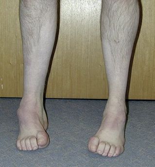
FIGURE 16.97
Classical appearance in the lower legs in a patient with Charcot-Marie-Tooth disease. D W Howcroft, S Kumar, N Makwana.
Orthopaedics and trauma
. 23(4):274–277, Fig 1. Elsevier, 2009, with permission.
3.
Acute intermittent porphyria.
4.
Diabetes mellitus.
5.
Lead poisoning.
6.
Multifocal motor neuropathy.
HINT
Motor neurone disease and neuromuscular junction disorders must always be considered in the differential diagnosis of distal motor weakness.
CAUSES OF A PREDOMINANTLY SENSORY NEUROPATHY (SENSORY NEURONOPATHY)
This is unusual and results in sensory ataxia and pseudoathetosis. Causes include the following:
1.
carcinoma (e.g. lung, ovary, breast)
2.
paraproteinaemia
3.
vitamin B
6
intoxication
4.
Sjögren’s syndrome
5.
diabetes mellitus
6.
syphilis
7.
vitamin B
12
deficiency (occasionally)
8.
idiopathic.
CAUSES OF A PAINFUL PERIPHERAL NEUROPATHY
1.
Diabetes mellitus.
2.
Alcohol.
3.
Vitamin B
12
or B
1
deficiency.
4.
Carcinoma.
5.
Porphyria.
6.
Arsenic or thallium poisoning.
7.
Heredity (most are not painful).
HINT
Burning soles of the feet can be caused by a painful peripheral neuropathy, tarsal tunnel syndrome or an S1 lesion. An S1 lesion will cause a decreased or absent ankle jerk.
CAUSES OF MONONEURITIS MULTIPLEX
Mononeuritis multiplex refers to separate involvement of more than one peripheral or rarely cranial nerve (e.g. a common peroneal nerve palsy plus an axillary nerve palsy). Common causes include:
1.
acute (usually vascular):
a.
diabetes mellitus
b.
polyarteritis nodosa or connective tissue diseases – SLE, rheumatoid arthritis.
2.
chronic:
a.
multiple compressive neuropathies, especially with joint-deforming arthritis
b.
sarcoidosis
c.
acromegaly
d.
leprosy
e.
Lyme disease
f.
carcinoma (rare)
g.
idiopathic.
CAUSES OF THICKENED NERVES
1.
Hereditary motor and sensory neuropathy.
2.
Acromegaly.
3.
Chronic inflammatory demyelinating polyradiculoneuropathy.
4.
Amyloidosis.
5.
Leprosy.
6.
Others – sarcoidosis, neurofibromatosis.
CAUSES OF FASCICULATION
Fasciculation is
not
always motor neurone disease. Causes include:
1.
benign idiopathic fasciculation (by far the most common)
2.
motor neurone disease
3.
motor root compression
4.
malignant neuropathy
5.
any motor neuropathy (less commonly).
HINT
Myokymia resembles benign coarse fasciculation of the same muscle group (e.g. eyelids). Electromyographic myokymia can occur in multiple sclerosis, brain stem neoplasm, Bell’s palsy, radiculopathy or radiation plexopathy or chronic nerve compression.
Hereditary motor and sensory neuropathy (HMSN)
Charcot-Marie-Tooth disease: usually autosomal dominant (see
Fig 16.96
).
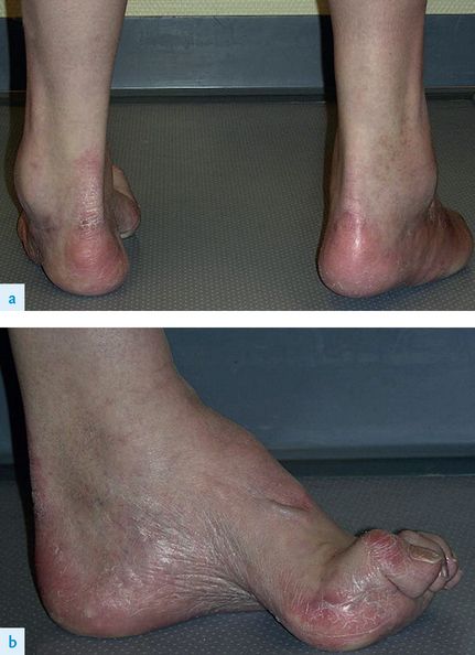
FIGURE 16.96
Typical clinical manifestations of a cavovarus foot deformity in a 55-year-old man who has CMT. This deformity is characterised by different components: (a) hindfoot varus and equinus (hind- or forefoot) and forefoot pronation, and (b) cavus, flexion deformity of the first metatarsal, and claw toes. T Dreher, S Hagmann, W Wenz. Reconstruction of multiplanar deformity of the hindfoot and midfoot with internal fixation techniques.
Foot and Ankle Clinics
, 2009. 14(3):489–531.
CLINICAL FEATURES
1.
Pes cavus (short, high-arched feet with hammer toes).
2.
Distal muscle atrophy owing to peripheral nerve degeneration, not usually extending above the elbows or above the middle one-third of the thighs.
3.
Absent reflexes.
4.
Slight to no sensory loss in the limbs.
5.
Thickened nerves.
6.
Optic atrophy; Argyll Robertson pupils (rare).
An approach to brachial plexus lesions
COMPLETE LESION
1.
Lower motor neurone signs affect the whole arm.
2.
Sensory loss (whole limb).
3.
Horner’s syndrome (an important clue, but only if the lesion is proximal in the lower plexus).
HINT
Remember always to feel for axillary lymphadenopathy at the end of your examination for a brachial plexus lesion.
UPPER TRUNK (ERB-DUCHENNE) (C5, C6) LESION
1.
Loss of shoulder movement and elbow flexion – hand is held in the ‘waiter’s tip’ position.
2.
Sensory loss is present over the lateral aspect of the arm and forearm, and over the thumb.
LOWER TRUNK (KLUMPKE) (C8, T1) LESION
1.
True claw hand with paralysis of all the intrinsic muscles.
2.
Sensory loss along the ulnar side of the hand and forearm.
3.
Horner’s syndrome.
CERVICAL RIB SYNDROME
1.
Weakness and wasting of the small muscles of the hand (true claw hand).
2.
Sensory loss over the medial aspect of the hand and forearm.
3.
Unequal radial pulses and blood pressures.
4.
Subclavian bruit and loss of the pulse on arm manoeuvring (this sign is often also present in normal persons).
5.
Palpable cervical rib in the neck (uncommon).
Important peripheral nerves
RADIAL NERVE (C5–C8) LESION
Clinical features
1.
Wrist and finger drop (wrist flexion normal).
2.
Triceps loss (elbow extension loss) if lesion is above the spiral groove.
3.
Sensory loss over the anatomical snuff box.
4.
Finger abduction
appears
to be weak because of the difficulty of spreading the fingers when they cannot be straightened.
MEDIAN NERVE (C6–T1) LESION
This nerve supplies all muscles on the front of the forearm except flexor carpi ulnaris and half of flexor digitorum profundus. It also supplies the following short muscles of the hand (LOAF):
L
ateral two lumbricals
O
pponens pollicis
A
bductor pollicis brevis
F
lexor pollicis brevis (this sometimes has ulnar innervation)
Clinical features
1.
Loss of abductor pollicis brevis with a lesion at or above the wrist – pen touching test: with the hand flat, ask the patient to abduct the thumb vertically to touch your pen.