Beyond the God Particle (24 page)
Read Beyond the God Particle Online
Authors: Leon M. Lederman,Christopher T. Hill
Tags: #Science, #Cosmology, #History, #Physics, #Nuclear, #General

Particle accelerators are, plainly and simply and precisely, the world's most powerful microscopes. To appreciate what these behemoths called particle accelerators are, let's take a look in detail at microscopes—let's put the microscope under the microscope.
MICROSCOPES
Ancients were aware of the phenomenon of lenses, but not much was done with them. The first serious practical application of lenses came with the invention of reading “spectacles” in the late thirteenth century, and primitive handheld “magnifiers” that were just tubes with a single lens at one end that could focus on a small object. An insect could be magnified by a few times, so these were often called “flea glasses.”
2
In 1590, two spectacle makers working in Holland (where they were probably counterfeiting coins), Hans Janssen and, particularly, his son Zacharias, discovered that with two lenses—one lens placed at each end of a tube—small objects could be made to appear greatly magnified.
3
This was the first
compound microscope
(several lenses), and it enabled magnification of small objects by about ten times. These early experiments also led to the telescope, which was perfected at about the same time, as was the science of optics, by Galileo. There is considerable uncertainty about the dates and attribution of these early developments of the microscope, involving a larger number of players (and no doubt considerable exaggeration and defamation), so we'll leave that to the historians. Certainly the
development of the microscope was intertwined with that of the telescope. The telescope had such immediate importance to seafaring navigation that the microscope seems to have emerged as a secondary spin-off, much like the World Wide Web was a spin-off of particle physics.
4
The celebrated “father of microscopy” and the “first microbiologist” was Anton Van Leeuwenhoek of Holland (1632–1723). Van Leeuwenhoek had no formal education, but he had considerable ingenuity and practical “street” skills. He apprenticed in a dry goods store where magnifying glasses were used to examine and count the number of threads in a fabric, and he later became a fabric merchant. Van Leeuwenhoek realized that high-quality lenses, with very short focal lengths, were needed to make better microscopes. This required extreme “double convex lenses,” that is, almost perfectly spherical little balls of glass. These type of lenses are much more of a challenge to make than are the larger, less curved lenses required for comparable magnifications for telescopes. These extreme lenses, crystal clear and perfectly spherical, also demanded a higher quality glass, instead of the greenish “coke-bottle” glass of the day. Van Leeuwenhoek began to make lenses of pure crystalline quartz, “painstakingly teaching himself and developing arduous new methods to grind and polish” these tiny, near-perfect spherical lenses. In actual fact, Van Leeuwenhoek had evidently discovered some cleverly simple methods of manipulating glass to achieve these lenses. He may have deliberately given the false impression that it was only through tedious and skillful grinding methods that he could achieve these results to stave off competitors:
Van Leeuwenhoek's interest in microscopes and a familiarity with glass processing led to one of the most significant technical insights in the history of science. By placing the middle of a small rod of glass in a hot flame, Van Leeuwenhoek could pull the melting section apart to create two long whiskers of glass. By reinserting the end of one whisker into the flame, he could create a very small glass sphere. These tiny spheres became the lenses of his microscopes, with the smallest spheres providing the highest magnifications. The shrewd Van Leeuwenhoek realized that if this simple method for making lenses was revealed, his role in microscopy might be minimized. He therefore allowed others to believe that he was laboriously spending most of his nights and free time grinding optically perfect tiny lenses to use in microscopes and he made about 200 high quality microscopes with different magnification powers.
5
Ultimately, he could achieve a whopping and unprecedented magnification up to 270 times. The modern microscope was born, and with it the science of microbiology. With this powerful new and revolutionary scientific instrument Van Leeuwenhoek's discoveries were bountiful. He was the first human to observe and describe bacteria (obtained from scrapings off of his teeth), yeast, and other tiny plants and microorganisms. He was first to discover the protozoan animal life in a drop of water from a pond and the circulation of blood cells in small capillaries. His many discoveries were published in over a hundred letters to the Royal Society of England and the French Academy. “My work, which I've done for a long time, was not pursued in order to gain the praise I now enjoy, but chiefly from a craving after knowledge, which I notice resides in me more than in most other men. And therewithal, whenever I found out anything remarkable, I have thought it my duty to put down my discovery on paper, so that all ingenious people might be informed thereof.”
6
Van Leeuwenhoek's scientific contemporary was the great English scientist Robert Hooke (a contender with Isaac Newton for the theory of gravitation).
7
Hooke had likewise improved on early compound microscopes around 1660 and had made a number of the key early discoveries. In his book “Micrographia” (1665), Hooke coined the word “cell” to describe the basic building blocks of the internal structure of the plant tissues that he and Van Leeuwenhoek were able to observe under their microscopes. Hooke benefited greatly from the work, and was a champion, of Van Leeuwenhoek.
Van Leeuwenhoek had discovered the single-celled animals, or protozoans. How remarkable to think of it, this grand moment of discovery of the biological cell and the tiniest organisms of life, the constituents of all higher organisms, much like the atom is constituent of all materials! Hooke promoted the scientific reputation and career of Van Leeuwenhoek and sought to have him installed in the Royal Society. Yet, in 1676, trouble developed between Van Leeuwenhoek and the Royal Society, trouble that paralleled Galileo's earlier difficulties with the Catholic Church over the observation of Jupiter's moons through a telescope. The validity of Van Leeuwenhoek's discovery in a drop of water of protozoans, the smallest single-celled animals, was vehemently challenged, the issue revolving
around evolution vs. religious preconceptions about life. Robert Hooke confirmed the discoveries, and only after considerable scrutiny and involvement of the English clergy was Van Leeuwenhoek ultimately vindicated in 1680. Observation and clear reason held the day, and Anton Van Leeuwenhoek became a Fellow of the Royal Society.
TECHNOLOGICAL CHALLENGE
The problem of making and using a nearly spherical lens is one of high technology, the highest of the late seventeenth century. A lens is always imperfect and creates a distorted image. One such distortion is the appearance of various fringes of color around boundaries in the image, called “chromatic aberration.” This occurs, not because of imperfections in the fabrication, but because different wavelengths of light are refracted by different amounts when they enter or exit a piece of glass.
8
The resulting details of an image appear splayed out like a rainbow, yielding the color-fringed image in both telescopes and microscopes. This is essentially the effect by which glass can serve as a prism for resolving white light into its constituent colors.
By the mid 1700s a clever solution was developed to this chromatic aberration problem in telescopes, the so-called “achromatic compound lens.” This is an arrangement of lenses that compensates the chromatic aberration and could be readily applied to the much less curved lenses in telescopes. But it remained a problem for the highly spherical microscope lens system. It was not until 1830 that Joseph Jackson Lister
9
(father of the famous surgeon Sir Joseph Lister, who was pioneer of antiseptic surgery) was able to make compound lenses to remedy the problem of chromatic aberration in microscopes.
Present-day optical microscopes are refinements of these early devices and give magnifications up to several thousand times. The perfection of high-quality-lens-based optical systems involves confronting and overcoming a number of various kinds of “aberrations.” Most of these are dealt with by exploiting the idea of compound lenses, where two or more lenses are placed in-line and mutually compensate each other's aberrations (this is analogous to how a married couple can be better behaved than each of the individuals separately!).
10
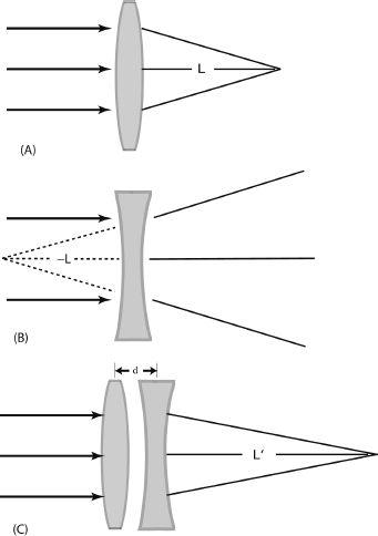
FIGURE 7.27. Lenses.
(A) A double convex lens has focal length L, the distance from the lens at which parallel incoming rays are focused; (B) a double concave lens causes parallel rays to diverge from a point and can be assigned a negative focal length –L; (C) combining together (A) and (B), with a small space between the lenses, d, yields a compound lens with a net focal length of L´ = L
2
/d, so the compound lens, which is known as “FODO” (focus-space-defocus-space) is “net focusing.” The compound lens can reduce aberrations of the two individual component lenses. This applies to magnetic lenses and forms the basic principle used in an “alternating gradient symnchrotron,” where repeating quadrupole magnets form an “…FODOFODO…” continuous focusing system, also called a “FODO lattice.”
The underlying idea of compound lenses involves a remarkable concept that turns out to be of foundational importance to the particle accelerators of the twentieth century. One can construct “convex” lenses that focus light to a point, modulo aberrations, and “concave” defocusing lenses that cause the beam or light to splay out. If a convex lens and a concave lens each having the same focal length are placed in-line close together but with a small air space between them,
the compound system is “net focusing
.
”
That is, the compound lens, consisting of a focusing-defocusing pair, will have a much longer focal length than either of the two lenses have separately. The net effect is that light in the compound system will always focus, or converge, to a point, even though the convex lens is defocusing (see
figure 7.27
). The aberrations due to imperfections of the two separate lenses may be about the same, but they will mostly cancel out in the compound lens system. This results in tolerably smaller aberrations, much smaller than for a single lens with the long focal length. As we'll see, this net focusing effect of a focusing-defocusing compound lens is the basis for the existence of large particle accelerators, called “synchrotrons,” such as the CERN LHC.
11
HOW DOES IT WORK?
Let's consider how a microscope system works in very general and simple terms. We start with a “beam of particles.” In the case of an optical microscope, this is a source of visible light. The particles are the
photons
that make up light (photons are quantum particles, so they behave both as particles and waves, but we'll put that aside for the moment). The source of the light, be it the sun or a candle or a lightbulb, is our “particle accelerator”—it has produced the particles, each of which carries energy as it moves toward us. We'll now put that beam to use in our microscope.
The microscope has a “target,” typically a glass slide upon which you place the thing you want to see, such as a drop of pond water that contains the protozoans you wish to observe or perhaps a section of your tonsils (after they were removed). The incoming photons of our particle beam collide with the target. In these collisions the beam particles are scattered in all directions.
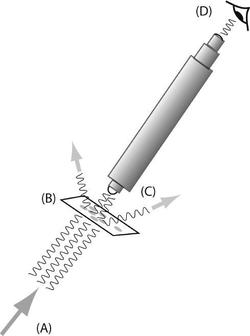
FIGURE 7.28. Microscope Schematic.
Schematic of a microscope (or of a particle accelerator experiment). The system has (A) an incoming beam of particles, such as photons; (B) a target off of which the particles scatter; (C) a detector that collects and processes the scattered particles and presents the data to (D) the human observer.