Examination Medicine: A Guide to Physician Training (38 page)
Read Examination Medicine: A Guide to Physician Training Online
Authors: Nicholas J. Talley,Simon O’connor
Tags: #Medical, #Internal Medicine, #Diagnosis

NASH is more common in women and the most frequently associated underlying clinical conditions are obesity, type 2 diabetes mellitus and hyperlipidaemia, particularly hypertriglyceridaemia. Look for evidence of the metabolic syndrome (including hypertension, increased waist circumference and high lipids). Nevertheless, studies have documented lean men with NASH. Although the peak age of presentation is the fifth and sixth decades of life, NASH is now the most common cause of liver disease in adolescents.
Exclude drug causes such as steroid use and amiodarone. The definitive diagnostic test is a liver biopsy but is not usually required. Other liver disease must be excluded (e.g. haemochromatosis, chronic viral hepatitis) (
Table 7.15
). The typical histological features are macrovesicular steatosis with an associated necroinflammatory infiltrate (usually mononuclear) and a variable degree of fibrosis.
Table 7.15
Causes of hepatic steatosis
| METABOLIC DISORDERS | DRUGS AND TOXINS |
| Obesity | Alcohol |
| Diabetes mellitus | Methotrexate |
| Hyperlipidaemia | Organic solvents |
| Jejunoileal bypass | Amiodarone |
| Total parenteral nutrition | |
Another group of patients has fatty liver disease without inflammation and with normal transaminase levels – non-alcoholic fatty liver disease (NAFLD).
A proportion of patients will progress to more chronic liver disease with fibrosis and ultimately cirrhosis. NASH may explain many cases previously labelled as cryptogenic. Studies have documented elevations in both cardiovascular and hepatic mortality.
Management
Management options are limited to treating the underlying clinical condition, with slow weight loss and exercise recommended for obese patients and strict control of hyperlipidaemia and hyperglycaemia. Statin therapy is safe for patients with hypercholesterolaemia. Alcohol should be avoided. Vitamin E supplementation is recommended in the absence of diabetes and if there is fibrosis.
Liver transplantation
Liver transplantation is now an important therapeutic option in patients with irreversible, progressive liver disease for which there is no acceptable alternative therapy and no absolute contraindication. The 1-year survival rate overall is 75%. In the examination setting, a patient will either have chronic liver disease and be a candidate for transplantation or be a transplant recipient who has a problem.
The history
1.
Obtain details of the patient’s liver disease, including diagnosis and duration. Candidates for liver transplantation include patients with cirrhosis, primary sclerosing cholangitis, autoimmune chronic hepatitis, chronic portal-systemic encephalopathy, Budd-Chiari syndrome, inherited metabolic diseases (e.g. Wilson’s disease, alpha
1
-antitrypsin deficiency) and acute or subacute hepatic failure.
2.
The timing of the transplantation is crucial. This should be considered when the patient is end-stage but before complications have occurred that may preclude proceeding (e.g. preterminal variceal bleeding, irreversible hepatorenal syndrome, development of a catabolic state, irreversible coagulopathy, vascular instability with ascites or incapacitating osteopenic bone disease). Those with end-stage liver disease who have had a life-threatening episode of decompensation or whose quality of life has become unbearably reduced are potential candidates. Accepted criteria include a Child-Pugh score >6, an episode of variceal bleeding or spontaneous bacterial peritonitis, or stage II encephalopathy in acute liver failure. A model for end-stage liver disease (MELD), which has been developed at the Mayo Clinic and is based on three parameters (serum bilirubin, INR and creatinine), helps predict survival and is widely used (transplant candidates have a score >10).
3.
If the patient may be a candidate for liver transplantation, enquire about potential contraindications. Relative contraindications include active sepsis outside the liver, metastatic malignancy, cholangiocarcinoma, continuing alcohol consumption, HIV, diffuse portal vein thrombosis and advanced cardiopulmonary or renal disease, a prior portacaval shunt (TIPS is not a contraindication), intrahepatic or biliary infection, localised portal vein thrombosis, renal failure, and prior complex hepatobiliary surgery. Severe hypoxaemia as a result of intrapulmonary shunting is another potential contraindication (patients with the hepatopulmonary syndrome and hypoxia can benefit, but a
Pa
O
2
of <50 mmHg is a relative contraindication; severe portopulmonary hypertension is a contraindication).
4.
Ask about tests that have been done in preparation for transplant. These typically include cardiac and pulmonary tests (electrocardiogram, echocardiogram, stress test, chest X-ray, pulmonary function tests), renal tests (urine protein and creatine, and glomerular filtration rate estimation) and liver tests (imaging to exclude a hepatoma and to define the vascular anatomy).
5.
Enquire about complications of the patient’s liver disease (e.g. previous variceal haemorrhages, ascites, pre-coma, hypoxaemia caused by hepatopulmonary syndrome etc.).
6.
If the patient has had a transplant, enquire about the postoperative course, including whether further surgery was carried out (e.g. drainage of abscesses, reconstruction of the biliary tract for control of bleeding, retransplantation for graft failure and for hepatic arterial thrombosis). Also ask about postoperative infections.
7.
Ask about complications of liver transplantation. Early on (in the first 5 days), primary graft failure, technical problems (e.g. bleeding, hepatic arterial thrombosis, bile leaks, portal vein thrombosis), renal failure and pulmonary complications (atelectasis, pleural effusion, infection) may occur. Major problems after discharge from hospital include rejection, infection, biliary complications, hypotension, recurrent disease, bone disease, nutrition and de-novo cancer. Infections occur in most patients, largely related to immunosuppression; chronic opportunistic infections usually occur from 4 weeks after transplantation. Biliary strictures may also occur from 4 weeks after transplantation. Acute liver rejection is rarely seen after the initial 6 months. Chronic rejection usually occurs 6 weeks to 9 months after transplantation; there is progressive cholestasis and diagnosis is best made by liver biopsy. Bone disease and ectopic calcification may occur some months after transplantation.
8.
Ask about current medications and related complications with their use. Cyclosporin may induce cholestasis (dose-dependent), hypertension, nephrotoxicity, gum hypertrophy, seizures (controlled by phenytoin, which itself induces cyclosporin metabolism) and central nervous system effects, including tremor and central pontine myelinolysis. Patients with a low serum cholesterol level are at increased risk of central nervous system toxicity. Steroids in high doses may induce a number of problems, including aseptic necrosis of long bones, cataracts and psychosis. Enquire about drug compliance.
9.
Ask about the specific complications of immunosuppression. Systemic and local infections are frequent problems and can be rapidly fatal if not treated aggressively. Opportunistic infections include
Pneumocystis jirovecii
and
Candida albicans
. Ask about prophylactic antibiotic therapy. Cytomegalovirus infection remains a major problem. Try to find out tactfully whether there has been any problem with malignancy. The incidence of skin cancers and lymphomas (especially in the central nervous system) is increased with immunosuppression.
The examination
1.
The pre-transplant patient should be examined for signs of chronic liver disease and complications of liver disease. Note clubbing (which occurs in cirrhosis and might indicate hepatopulmonary syndrome is this setting).
2.
The post-transplant patient should be examined for liver tenderness (e.g. acute rejection) and jaundice (e.g. vanishing bile ducts in chronic rejection, biliary stricture). Examine the chest for infection and the mouth for candidiasis. The temperature must be taken (
Table 7.16
). Examine the central nervous system (e.g. cyclosporin toxicity or cerebral infarction from perioperative hypotension or air embolism). Tap the spine for tenderness (e.g. vertebral collapse). Take the blood pressure (hypertension may occur at any time after transplantation; it is often caused by cyclosporin).
Table 7.16
Causes of fever in the outpatient with a liver transplant
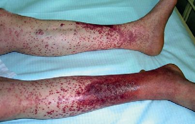
FIGURE 7.13
Mixed cryoglobulinaemia in a patient with hepatitis C. Purpura of the legs. M Ramos-Casals. The cryoglobulinaemias.
The Lancet
. 2012. 379(9813):348–360. Elsevier, with permission.
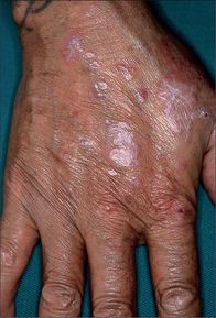
FIGURE 7.14
Lichen planus on the dorsal surface of the hand. Wickham’s striae can be easily identified in the upper right lesion. Note the flat-topped nature of the lesions. J L Bolognia, J L Jorizzo, J V Schaffer.
Dermatology
. 3rd edn. Elsevier, 2012, with permission.
Biliary tract (stricture and cholangitis)
Pneumonia (e.g.
Pneumocystis
, bacterial, fungal)
Urinary tract sepsis
Hepatitis (acute or recurrent)
Central nervous system infection (especially fungal)
Viral infection (e.g. cytomegalovirus, herpes, varicella-zoster)
Investigations
Pre-transplant patients need tests to confirm the diagnosis, determine current liver synthetic function (e.g. serum albumin, INR) and rule out contraindications. Ultrasound and CT scanning are routine. In patients with possible or definite malignancy, metastases must be sought. Assess bone density; osteopenia is common in liver disease and increases following transplant. Cardiopulmonary and psychiatric evaluation are important.
Management
Routine outpatient monitoring after transplantation should include a full blood count, electrolyte levels, renal and liver profile, and trough cyclosporin levels. Remember drug interactions with cyclosporin. Diabetes mellitus may supervene as a result of treatment with tacrolimus or steroids. Hypertension should be treated. Diseases that can recur in the graft include hepatitis B and C, Budd-Chiari syndrome and primary biliary cirrhosis.
CHAPTER 8
The haematological long case
The modern haematologist, instead of describing in English what he can see, prefers to describe in Greek what he can’t.
Richard Allan John Asher (1912–69)
Haemolytic anaemia
This is an uncommon but important long case. It is usually a diagnostic problem. Coombs’ positive haemolytic anaemia is most often encountered in the examination.
The history
1.
Ask about the symptoms of anaemia (e.g. fatigue, shortness of breath on exertion) and whether the patient has noticed or been told about jaundice.
2.
Determine whether there is a history of known haemolytic episodes. Onset at an early age or a family history suggests an intrinsic red cell defect (e.g. hereditary spherocytosis or elliptocytosis (both autosomal dominant), sickle cell anaemia).

